

An ultrasound scan is one of the most commonly performed diagnostic imaging tests in modern healthcare. It helps doctors examine internal organs, monitor pregnancy, evaluate blood flow, and diagnose a wide range of medical conditions without using radiation. Since it is safe, painless, and non-invasive, ultrasound is often the first imaging test recommended for many health concerns.
Whether you need an ultrasound for pregnancy, abdominal pain, breast evaluation, or another medical condition, choosing a trusted diagnostic center is essential for accurate results. To book an ultrasound scan or speak with our team, contact MVR Diagnostic Clinic.
Read on to learn how ultrasound scans work, the different types available, what they can detect, and how to prepare for your appointment.
An ultrasound scan, also known as ultrasonography or sonography, is a medical imaging test that uses high-frequency sound waves to produce real-time images of organs, tissues, muscles, blood vessels, and other structures inside the body.
Unlike X-rays and CT scans, ultrasound does not use ionising radiation. Instead, it relies on harmless sound waves, making it a safe imaging method for people of all ages, including pregnant women and babies.
Doctors use ultrasound scans to diagnose medical conditions, monitor pregnancy, guide certain medical procedures, and evaluate how different organs are functioning.
Because ultrasound produces images in real time, healthcare providers can observe organs and tissues while they are moving, providing valuable diagnostic information that cannot always be obtained with other imaging techniques.
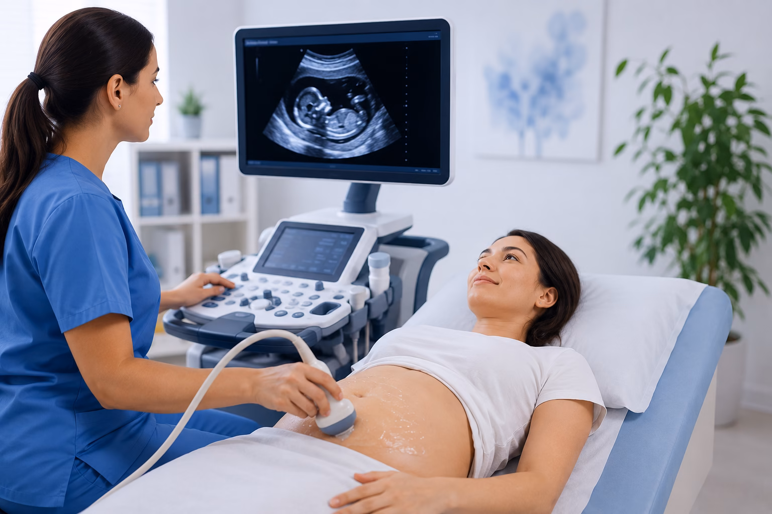
Ultrasound technology works by sending high-frequency sound waves into the body. These sound waves bounce off internal organs and tissues, creating echoes that are converted into detailed images by a computer. The entire process is painless and usually takes between 15 and 45 minutes, depending on the type of examination being performed.
An ultrasound machine consists of a computer, monitor, and a handheld device called a transducer. The transducer sends sound waves into the body and receives the returning echoes. To improve contact with the skin, a clear water-based gel is applied before the examination begins. This allows the sound waves to travel efficiently and produce clearer images.
One of the biggest advantages of ultrasound is its ability to display images instantly. As the transducer moves over the body, doctors can observe organs, blood vessels, muscles, and even a developing baby in real time. This makes ultrasound particularly useful for monitoring movement, guiding biopsies, and assessing blood circulation.
Unlike CT scans and X-rays, ultrasound relies entirely on sound waves rather than radiation. This makes it one of the safest diagnostic imaging techniques available. Since there is no radiation exposure, ultrasound is widely used throughout pregnancy and can be repeated whenever medically necessary without the risks associated with ionising radiation.
Ultrasound scans help doctors diagnose, monitor, and evaluate a wide variety of health conditions. Depending on the area being examined, the scan provides valuable information about organs, blood vessels, muscles, and developing babies during pregnancy.
Ultrasound is best known for monitoring pregnancy. It allows doctors to confirm pregnancy, estimate the due date, monitor fetal growth, assess the baby’s heartbeat, evaluate the placenta, and detect certain developmental abnormalities.
If you are planning a pregnancy ultrasound, you may also find our guide on Pregnancy Ultrasound Scan Cost in Dubai helpful. It explains the different types of pregnancy scans and what to expect during each stage.
Ultrasound is commonly used to examine several internal organs, including:
It helps detect inflammation, cysts, tumours, stones, infections, and other abnormalities affecting these organs.
Doctors frequently recommend ultrasound for patients experiencing abdominal or pelvic pain. The examination can identify conditions such as gallstones, kidney stones, ovarian cysts, uterine fibroids, enlarged organs, and fluid accumulation. Pelvic ultrasound is also used to evaluate the uterus, ovaries, prostate, and urinary bladder.
Breast ultrasound is often performed to investigate breast lumps, breast pain, or abnormalities detected during a mammogram. It helps distinguish fluid-filled cysts from solid masses and provides additional information for diagnosis.
Different types of ultrasound scans are used depending on the part of the body being examined. Each scan provides detailed images that help doctors diagnose specific medical conditions.
An abdominal ultrasound examines organs within the abdomen, including the liver, gallbladder, kidneys, pancreas, and spleen. It is commonly recommended for abdominal pain, suspected gallstones, kidney stones, liver disease, and digestive problems.
A pelvic ultrasound evaluates the reproductive organs and bladder. In women, it helps assess the uterus, ovaries, and cervix, while in men it may be used to examine the prostate and bladder. This scan is often performed to investigate pelvic pain, abnormal bleeding, infertility, ovarian cysts, or uterine fibroids.
A transvaginal ultrasound provides detailed images of the female reproductive organs using a specially designed probe inserted into the vagina. Doctors commonly use this examination during early pregnancy and to evaluate fertility concerns, pelvic pain, endometriosis, ovarian cysts, and uterine abnormalities.
A breast ultrasound creates detailed images of breast tissue and is frequently used to evaluate breast lumps, nipple discharge, breast pain, and abnormalities found during mammography. It is particularly valuable for women with dense breast tissue.
A thyroid ultrasound examines the thyroid gland located in the neck. It helps identify thyroid nodules, cysts, enlargement, inflammation, and other thyroid disorders.
Pregnancy ultrasounds monitor the baby’s growth and development throughout pregnancy. Different scans are performed at various stages to assess fetal health, measure growth, examine anatomy, and monitor the placenta and amniotic fluid.
Many parents also wonder when an ultrasound can accurately determine their baby’s gender. If you’re curious about this, read our guide on How Accurate Is an Early Gender Ultrasound Scan During Pregnancy?.
A 4D ultrasound creates moving three-dimensional images of the baby in real time. It allows parents and doctors to observe facial expressions, movements, and other developmental features during pregnancy. Although often performed for bonding purposes, 4D ultrasound can also provide additional clinical information in selected cases.
A Doppler ultrasound evaluates blood flow through arteries and veins. It helps detect blocked blood vessels, blood clots, narrowed arteries, poor circulation, and other vascular conditions. Doctors also use Doppler ultrasound during pregnancy to monitor blood flow between the placenta and the developing baby.
Ultrasound scans are used to detect a wide range of medical conditions affecting different organs and tissues throughout the body. Since the scan provides real-time images, it allows doctors to identify abnormalities quickly and accurately without exposing patients to radiation. Depending on the area being examined, an ultrasound scan can detect the following conditions.
Ultrasound is one of the most common imaging tests used to diagnose gallstones and kidney stones. It can identify the size, location, and number of stones, helping doctors determine the most appropriate treatment. Patients experiencing severe abdominal pain, flank pain, or urinary symptoms are often referred for an ultrasound examination.
Ultrasound can detect both solid masses and fluid-filled cysts in organs such as the liver, kidneys, thyroid, ovaries, breasts, and other soft tissues. Although ultrasound helps identify these abnormalities, additional imaging or a biopsy may sometimes be required to confirm the diagnosis.
During pregnancy, ultrasound helps monitor both the mother and the baby. It can identify multiple pregnancies, ectopic pregnancy, placenta-related problems, reduced amniotic fluid, fetal growth concerns, and certain congenital abnormalities. Regular pregnancy ultrasounds help ensure the healthy development of the baby throughout each trimester.
Inflammation affecting organs such as the liver, gallbladder, pancreas, kidneys, and thyroid can often be detected using ultrasound. The examination helps doctors diagnose infections, swelling, enlarged organs, and other inflammatory conditions that may require medical treatment.
A Doppler ultrasound evaluates blood circulation through arteries and veins. It can detect narrowed blood vessels, blocked arteries, blood clots, varicose veins, and reduced blood flow to different parts of the body.
Ultrasound plays an essential role throughout pregnancy by monitoring fetal development and identifying any concerns that may require medical attention. Different ultrasound scans are performed at various stages of pregnancy, each serving a specific purpose.
The dating scan is usually performed during the first trimester to confirm pregnancy, estimate the baby’s due date, determine the number of babies, and assess the baby’s heartbeat. Accurate pregnancy dating helps doctors plan future prenatal care.
The NT scan is typically performed between 11 and 13 weeks of pregnancy. It measures the fluid at the back of the baby’s neck and is commonly combined with blood tests to assess the risk of certain chromosomal conditions.
Also known as the anomaly scan, this detailed examination is usually performed between 18 and 22 weeks of pregnancy. It evaluates the baby’s organs, brain, heart, spine, limbs, kidneys, face, and other anatomical structures while also examining the placenta and amniotic fluid.
A growth scan is usually performed during the later stages of pregnancy to monitor fetal growth, estimate the baby’s weight, assess amniotic fluid levels, and evaluate placental function. This scan helps ensure that the baby continues to grow as expected.
Throughout pregnancy, ultrasound allows doctors to monitor the baby’s heartbeat, movements, breathing activity, growth, and overall well-being. These routine examinations provide reassurance for expectant parents while helping healthcare providers identify any complications at an early stage.
Preparation for an ultrasound scan depends on the type of examination being performed. Following the correct instructions helps produce clearer images and improves diagnostic accuracy.
Some ultrasound examinations, particularly abdominal scans, require fasting for several hours before the appointment. An empty stomach helps reduce bowel gas and allows better visualisation of organs such as the liver, gallbladder, and pancreas. Always follow the instructions provided by your healthcare provider.
For pelvic ultrasound examinations, patients are often asked to drink several glasses of water before the scan and avoid emptying their bladder. A full bladder improves image quality by providing a clearer view of the pelvic organs.
To make your appointment more comfortable:
Most ultrasound examinations are painless and can be completed within 15 to 45 minutes.
Different imaging techniques provide different types of diagnostic information. Your doctor will recommend the most appropriate examination based on your symptoms and medical condition.
A CT scan uses X-rays to create detailed cross-sectional images of the body. It is particularly useful for evaluating bones, lungs, internal injuries, and certain cancers.
Ultrasound, on the other hand, uses sound waves instead of radiation. It is commonly used for pregnancy, abdominal organs, soft tissues, blood vessels, and breast imaging.
While CT scans provide highly detailed images of many internal structures, ultrasound is often preferred when radiation exposure should be avoided.
MRI uses powerful magnets and radio waves to create highly detailed images of soft tissues, joints, muscles, the brain, and internal organs.
Ultrasound is generally quicker, more affordable, and provides real-time imaging. MRI offers greater detail for complex medical conditions but usually requires a longer examination.
Your healthcare provider will recommend the imaging test that best answers your specific medical question.
Ultrasound is one of the safest imaging methods available because it does not expose patients to radiation. Its accuracy depends on several factors, including the area being examined, the patient’s body type, and the experience of the sonographer. In many situations, ultrasound is combined with CT or MRI to provide a more comprehensive diagnosis.
Ultrasound offers several important benefits, making it one of the most frequently performed diagnostic imaging tests worldwide.
Unlike X-rays and CT scans, ultrasound uses sound waves instead of ionising radiation, making it suitable for pregnant women, children, and patients requiring repeated imaging.
Most ultrasound examinations can be completed within a short time, allowing doctors to evaluate the images immediately and prepare diagnostic reports promptly.
Compared with many other imaging techniques, ultrasound is generally more affordable while still providing valuable diagnostic information for a wide range of medical conditions.
Ultrasound has been safely used in pregnancy for many years and remains the preferred imaging method for monitoring fetal development without exposing the baby to radiation.
Although ultrasound is highly valuable, it also has certain limitations. Understanding these helps explain why doctors sometimes recommend additional imaging tests.
Ultrasound cannot clearly visualise structures hidden behind bone or organs containing large amounts of air, such as the lungs and parts of the digestive tract. For these areas, CT or MRI may provide better diagnostic information.
Image quality can sometimes be affected by obesity, excessive bowel gas, or the location of the organ being examined. In such situations, additional imaging may be recommended.
The quality of an ultrasound examination depends on the experience and expertise of the sonographer performing the scan and the radiologist interpreting the images. For this reason, choosing an experienced diagnostic centre is important for obtaining accurate results.
At MVR Diagnostic Clinic, we provide comprehensive ultrasound services using advanced imaging technology to help diagnose and monitor a wide range of medical conditions. Our experienced radiologists and sonographers perform high-quality ultrasound examinations with a strong focus on patient comfort, safety, and accurate diagnosis. Whether you require a pregnancy ultrasound, abdominal scan, breast ultrasound, thyroid ultrasound, pelvic ultrasound, or Doppler study, our team is committed to delivering reliable diagnostic imaging and timely reports to support your healthcare journey.
To schedule your ultrasound scan or learn more about our services, contact MVR Diagnostic Clinic. Our team will be happy to assist you with appointments and answer any questions about your ultrasound examination.
Ultrasound scans have become one of the most valuable diagnostic tools in modern medicine. They provide real-time images of internal organs and tissues, helping doctors identify medical conditions quickly without exposing patients to radiation. From monitoring a healthy pregnancy to diagnosing abdominal pain, evaluating breast lumps, or assessing blood flow, ultrasound supports accurate diagnosis across many areas of healthcare.
Since every medical condition is unique, your doctor will recommend the most appropriate type of ultrasound based on your symptoms and medical history. Following the recommended preparation instructions and choosing an experienced diagnostic centre helps ensure clear images and reliable results.
If your healthcare provider recommends an ultrasound scan, timely imaging can lead to earlier diagnosis, appropriate treatment, and greater peace of mind. At MVR Diagnostic Clinic, we are committed to providing accurate, compassionate, and patient-focused diagnostic care for individuals and families across the UAE.
What is an ultrasound scan used for?
An ultrasound scan is used to examine internal organs, monitor pregnancy, evaluate blood vessels, investigate pain or swelling, and diagnose a wide range of medical conditions affecting different parts of the body.
Is an ultrasound scan painful?
No. Ultrasound is a painless and non-invasive imaging test. You may feel slight pressure as the transducer moves over the skin, but the procedure is generally comfortable.
Does an ultrasound scan use radiation?
No. Ultrasound uses high-frequency sound waves rather than radiation, making it a safe imaging option for adults, children, and pregnant women.
How long does an ultrasound scan take?
Most ultrasound examinations take between 15 and 45 minutes. The exact duration depends on the type of scan and the body part being examined.
Do I need to fast before an ultrasound scan?
Some abdominal ultrasound scans require fasting for several hours before the examination. However, many other ultrasound scans do not require fasting. Your healthcare provider will give you specific preparation instructions.
Why do I need a full bladder for a pelvic ultrasound?
A full bladder helps lift the intestines away from the pelvic organs, providing clearer images of the uterus, ovaries, bladder, and surrounding structures.
Can an ultrasound detect cancer?
Ultrasound can identify abnormal masses, cysts, or tumours that may require further investigation. However, additional imaging or a biopsy is often needed to confirm whether an abnormality is cancerous.
Is ultrasound safe during pregnancy?
Yes. Ultrasound has been safely used during pregnancy for many years. It is the preferred imaging method for monitoring fetal growth and development because it does not expose the mother or baby to radiation.
What should I wear for an ultrasound appointment?
Wear loose, comfortable clothing that allows easy access to the area being examined. Depending on the type of scan, you may be asked to change into a medical gown.
How soon will I receive my ultrasound results?
The images are reviewed by a radiologist, who prepares a detailed report for your referring doctor. The time required to receive results varies depending on the examination and the healthcare facility.
WhatsApp us