

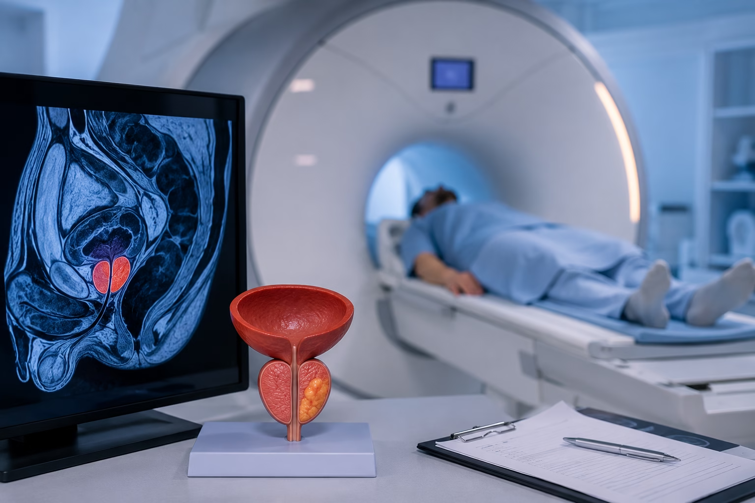
Prostate cancer is one of the most common cancers affecting men worldwide, and early detection plays an important role in choosing the most appropriate treatment. While blood tests such as the prostate-specific antigen (PSA) test can indicate that further investigation may be needed, they cannot confirm whether cancer is present. This is where a prostate MRI becomes an important part of the diagnostic process.
Modern MRI technology provides detailed images of the prostate, helping doctors identify suspicious areas, assess the extent of disease, and determine whether a biopsy is necessary. Whether your doctor has recommended an MRI because of an elevated PSA level, an abnormal examination, or to monitor an existing condition, understanding the procedure can help you feel more prepared. Read on to learn how a prostate MRI works, when it is recommended, how to prepare, and what your results may mean.
A prostate MRI is a specialised imaging examination that uses a powerful magnetic field and radio waves to produce highly detailed images of the prostate gland and surrounding tissues. Unlike X-rays or CT scans, MRI does not use ionising radiation, making it a safe imaging option for many patients.
The prostate is a small gland located below the bladder and in front of the rectum. It produces fluid that forms part of semen and plays an important role in male reproductive health. Because the prostate is surrounded by other delicate structures, MRI provides excellent soft tissue detail, allowing doctors to examine the gland more accurately than many other imaging techniques.
A prostate MRI may help detect abnormalities such as:
Depending on your medical history and clinical findings, your doctor may recommend a standard prostate MRI or a more advanced multiparametric MRI (mpMRI) to obtain additional diagnostic information.
If you’re unfamiliar with MRI scans, our MRI Scan: Complete Guide to Procedure, Preparation, Uses & Results explains how MRI technology works and what you can expect during your examination.
A prostate MRI helps doctors evaluate abnormalities that cannot always be identified through physical examination or blood tests alone. It provides detailed information that supports diagnosis, treatment planning, and ongoing monitoring.
Common reasons for requesting a prostate MRI include:
Elevated PSA Levels
One of the most common reasons for a prostate MRI is a raised prostate-specific antigen (PSA) level. Although an elevated PSA does not necessarily mean cancer is present, it may indicate that additional investigation is required.
To understand how blood tests such as PSA contribute to health screening, read our Blood Test Guide: Types, Results, Normal Ranges & Common Tests.
Abnormal Digital Rectal Examination (DRE)
If your doctor detects an unusual area during a digital rectal examination, an MRI can provide detailed images to determine whether further investigation is needed.
Suspected Prostate Cancer
MRI is frequently used when prostate cancer is suspected based on symptoms, PSA results, or clinical examination.
Before a Prostate Biopsy
Many specialists now recommend MRI before performing a biopsy. MRI can identify suspicious areas within the prostate, allowing biopsies to target the most concerning regions rather than sampling the gland randomly.
After a Previous Negative Biopsy
Sometimes a biopsy may not detect cancer even though PSA levels remain elevated. MRI can help identify areas that may have been missed during earlier testing.
Active Surveillance
Men diagnosed with low-risk prostate cancer may undergo regular MRI scans to monitor changes over time and determine whether treatment is necessary.
Cancer Staging
Once prostate cancer has been diagnosed, MRI helps evaluate whether the disease is confined to the prostate or has spread into nearby tissues.
Treatment Planning
Detailed MRI images assist healthcare providers in selecting the most appropriate treatment approach, whether surgery, radiation therapy, or active surveillance.
Monitoring After Treatment
MRI may also be used to assess treatment response or investigate signs of cancer recurrence following surgery or other therapies.
To learn how a prostate MRI helps detect and evaluate prostate cancer, read MRI for Prostate Cancer Detection.
Multiparametric MRI, commonly known as mpMRI, is an advanced form of prostate MRI that combines several imaging techniques to provide a more detailed assessment of the prostate gland.
Rather than relying on a single type of image, mpMRI evaluates different characteristics of prostate tissue, helping radiologists distinguish between normal tissue and areas that may require further investigation.
An mpMRI typically includes:
T2-Weighted Imaging
This sequence produces highly detailed anatomical images of the prostate and helps identify structural abnormalities within the gland.
Diffusion-Weighted Imaging (DWI)
Diffusion-weighted imaging measures the movement of water molecules within tissues. Cancer cells often restrict this movement, making suspicious areas easier to identify.To understand how diffusion MRI supports early cancer detection, read Why Diffusion MRI Is Changing the Way Cancer Is Detected Early.
Dynamic Contrast-Enhanced Imaging (DCE)
For some examinations, a contrast agent is injected into a vein to evaluate blood flow within the prostate. Areas with abnormal blood supply may warrant closer evaluation.
Together, these imaging techniques provide a comprehensive assessment of the prostate and improve the detection of clinically significant cancers while helping reduce unnecessary biopsies.
Today, multiparametric MRI is widely regarded as the preferred imaging method for evaluating patients with suspected prostate cancer.
Preparing for a prostate MRI is simple, but following the recommended instructions helps ensure high-quality images and a smooth examination.
Before your appointment:
If you’re having your first MRI, our MRI Preparation Guide: What to Expect Before Your Scan explains how to prepare and what happens during the examination.
On the day of your appointment, a radiographer will explain the procedure and answer any questions before the scan begins.
During the examination:
Most prostate MRI examinations take approximately 30 to 45 minutes, although this may vary depending on the imaging protocol used.
A prostate MRI is generally not painful.
The examination is completely non-invasive, and most patients experience little or no discomfort while lying on the scanning table.
Some people may find it slightly uncomfortable to remain still for the duration of the scan, particularly if they have back pain or joint stiffness. If contrast material is used, you may feel a brief pinch when the intravenous cannula is inserted.
Patients who feel anxious in enclosed spaces should let the imaging team know before the appointment. Modern wide-bore MRI scanners provide a more spacious environment, helping improve comfort for many patients.
MRI has become one of the most valuable imaging tools for evaluating prostate disease because it provides excellent visualisation of soft tissues without exposing patients to ionising radiation.
Some of the key benefits include:
Improved Detection of Significant Prostate Cancer
MRI helps identify suspicious areas that may represent clinically significant prostate cancer while reducing the detection of cancers that are unlikely to require immediate treatment.
Helps Guide Targeted Biopsies
Rather than performing random tissue sampling, MRI allows doctors to target suspicious lesions more accurately during biopsy.
Supports Accurate Cancer Staging
MRI helps determine whether cancer is confined to the prostate or has spread into surrounding tissues, supporting treatment planning.
Useful for Active Surveillance
Patients with low-risk prostate cancer may undergo regular MRI examinations to monitor disease progression and determine whether treatment is necessary.
No Radiation Exposure
Unlike CT scans, MRI uses magnetic fields and radio waves instead of ionising radiation.
High Soft Tissue Detail
MRI produces highly detailed images that help radiologists evaluate the prostate and nearby structures with excellent clarity.
Although prostate MRI is an excellent diagnostic tool, it also has some limitations.
These include:
For this reason, MRI findings are always interpreted alongside PSA levels, physical examination, biopsy results, and your overall medical history.
After your examination, an experienced radiologist reviews the MRI images and prepares a detailed report for your referring doctor.
Depending on the findings, your report may indicate:
Normal Appearance
The prostate appears normal, with no suspicious abnormalities identified.
Benign Changes
The scan may show non-cancerous conditions such as benign prostatic enlargement (BPH) or prostatitis.
Suspicious Lesions
If an abnormal area is detected, the report will usually include a PI-RADS score to describe the likelihood of clinically significant prostate cancer.
Recommendation for Further Investigation
If the MRI identifies a suspicious lesion, your doctor may recommend a targeted prostate biopsy to confirm the diagnosis.
Your healthcare provider will explain the MRI findings alongside your PSA level, symptoms, physical examination, and any previous investigations before recommending the next steps.
Choosing an experienced diagnostic centre is essential for obtaining high-quality prostate MRI images and accurate interpretation.
At MVR Diagnostic Clinic, patients benefit from advanced imaging technology and expert care throughout the diagnostic process.
Advanced MRI Technology
Our 1.5 Tesla Siemens Wide-Bore MRI with a 70 cm bore provides excellent image quality while offering a more comfortable experience for many patients.
Experienced Radiologists
Every MRI examination is reviewed by experienced radiologists who provide accurate, detailed reports to support diagnosis and treatment planning.
Comfortable Scanning Environment
Our wide-bore MRI system helps reduce discomfort for many patients, including those who experience anxiety or mild claustrophobia.
Convenient Appointments
We offer flexible scheduling to help patients receive timely diagnostic imaging when they need it.
Patient-Centred Care
Our team is available to answer your questions, explain the procedure, and guide you through every stage of your MRI examination.
A prostate MRI is an important tool for evaluating prostate abnormalities, assessing the likelihood of clinically significant cancer, and supporting treatment decisions. Combined with PSA testing, clinical examination, and, when necessary, biopsy, MRI provides valuable information that helps doctors deliver more personalised care.
If your doctor has recommended a prostate MRI or you’re concerned about your prostate health, early evaluation can provide reassurance and help identify problems before they become more advanced.
Book your prostate MRI at MVR Diagnostic Clinic and benefit from advanced 1.5 Tesla Siemens Wide-Bore MRI technology, experienced radiologists, and compassionate patient care.
What is a prostate MRI?
A prostate MRI is an imaging examination that uses magnetic fields and radio waves to produce detailed images of the prostate gland and surrounding tissues.
Can MRI detect prostate cancer?
MRI can identify suspicious areas that may indicate clinically significant prostate cancer, but a biopsy is usually required to confirm the diagnosis.
What is a PI-RADS score?
PI-RADS is a standardised scoring system that estimates how likely a prostate lesion is to represent clinically significant prostate cancer, using a scale from 1 to 5.
How long does a prostate MRI take?
Most prostate MRI examinations take approximately 30 to 45 minutes.
Will I need contrast?
Some prostate MRI scans include contrast material to improve image quality. Your healthcare provider will determine whether contrast is appropriate for your examination.
Is a prostate MRI painful?
No. A prostate MRI is generally painless, although remaining still for the duration of the scan may cause mild discomfort for some patients.
Can MRI replace a prostate biopsy?
MRI is an excellent diagnostic tool, but it cannot confirm cancer on its own. If suspicious findings are present, a biopsy may still be required.
How accurate is a prostate MRI?
Modern multiparametric MRI is highly accurate for detecting clinically significant prostate cancer and plays an important role in diagnosis, treatment planning, and active surveillance.
Who should have a prostate MRI?
Your doctor may recommend a prostate MRI if you have elevated PSA levels, an abnormal digital rectal examination, suspected prostate cancer, or require monitoring after diagnosis or treatment.
How should I prepare for a prostate MRI?
Follow your imaging centre’s instructions, remove all metal objects, inform staff about implants or medical devices, and complete any preparation advised before your appointment.
WhatsApp us