

Early detection plays a vital role in improving cancer treatment and survival rates. The sooner cancer is identified, the more treatment options are available and the better the chances of successful outcomes. Advances in medical imaging have made it possible to detect certain cancers much earlier than before, helping doctors make faster and more accurate diagnoses.
One of these advancements is Diffusion MRI. This specialized MRI technique provides valuable information about how water molecules move within body tissues, allowing doctors to identify abnormal cellular changes that may not be visible on conventional imaging. It has become an important tool for detecting cancer, assessing tumour characteristics, and monitoring treatment response.
Read on to learn how Diffusion MRI works, why it is different from a standard MRI, and how it is helping transform the early detection of cancer.
Diffusion MRI, also known as Diffusion-Weighted Imaging (DWI), is an advanced MRI technique that measures the movement of water molecules within the body’s tissues. Healthy and diseased tissues affect water movement differently, allowing radiologists to identify abnormalities that may indicate the presence of cancer or other medical conditions.
Unlike imaging tests that mainly show the structure of organs, Diffusion MRI provides functional information about tissues at the cellular level. This allows doctors to detect certain diseases before visible structural changes develop.
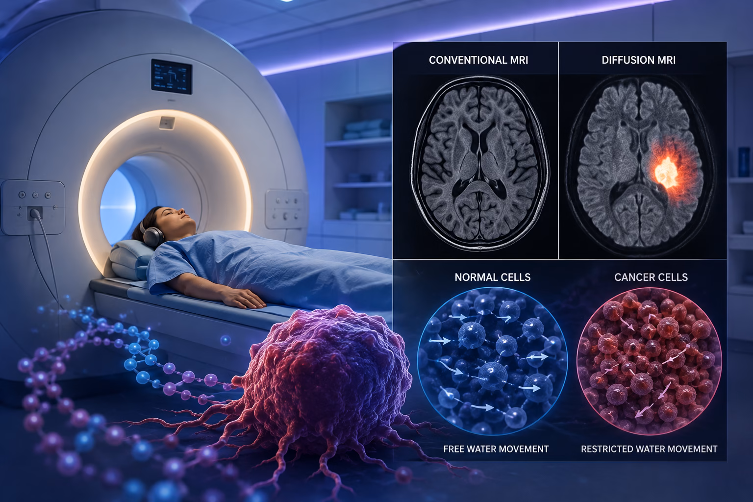
Water molecules are constantly moving inside the body’s cells and tissues. In healthy tissue, water moves relatively freely. However, cancer cells are usually packed closely together, restricting the movement of water molecules.
Diffusion MRI measures these differences in water movement and converts them into detailed images. Areas where water movement is restricted often appear brighter on the scan, helping radiologists identify suspicious tissue that may require further evaluation.
To better understand how diffusion-weighted imaging works and why it plays a crucial role in modern MRI, read Diffusion Weighted Imaging: All You Need to Know.
A conventional MRI creates detailed pictures of organs, muscles, bones, and soft tissues. While these images show the size, shape, and structure of different body parts, they may not always reveal subtle cellular changes.
Diffusion MRI goes one step further by examining tissue function rather than structure alone. It can detect microscopic changes that may occur before a tumour becomes clearly visible on a routine MRI scan.
For many cancers, combining conventional MRI with diffusion imaging provides a more complete picture and improves diagnostic accuracy.
For a detailed comparison of diffusion MRI and standard MRI, read DWI MRI Brain vs Regular MRI: What You Should Know Before Choosing a Scan.
Is Diffusion MRI Safe?
Yes. Diffusion MRI is considered a safe imaging technique because it uses powerful magnets and radio waves instead of ionising radiation.
The examination is non-invasive and painless. In many cases, diffusion imaging can be performed as part of a routine MRI scan without requiring any additional procedures. Some MRI examinations may still require contrast material depending on the body part being examined and the clinical question.
Cancer often develops gradually, and symptoms may not appear until the disease has progressed. Detecting cancer during its early stages gives doctors a better opportunity to begin treatment before it spreads to nearby tissues or other parts of the body.
Early diagnosis offers several important benefits.
When cancer is detected early, treatment is generally more effective. Patients diagnosed at an early stage often have a greater chance of successful treatment and long-term recovery.
Early-stage cancers can often be treated using less aggressive methods. Depending on the type of cancer, patients may have access to surgery, targeted therapies, or localized treatments before the disease advances.
Many cancers have significantly higher survival rates when diagnosed early. Regular screening and advanced imaging techniques help identify abnormalities before symptoms become severe.
Smaller tumours usually require less complex treatment than advanced cancers. Early detection may reduce the need for extensive surgery, intensive chemotherapy, or radiation therapy.
Receiving treatment before cancer progresses often allows patients to recover more quickly and maintain a better quality of life during and after treatment.
Diffusion MRI has become an important part of modern cancer imaging because it can identify changes within tissues before they become obvious on conventional imaging. By evaluating how water molecules move inside cells, this technique helps doctors detect suspicious areas with greater confidence.
Cancer begins with abnormal changes inside cells. Diffusion MRI can identify these microscopic changes before noticeable structural changes appear on routine imaging, making early diagnosis more likely.
Cancer cells are usually more densely packed than healthy cells. This restricts the normal movement of water molecules within the tissue.
Diffusion MRI measures these differences, helping radiologists distinguish abnormal tissue from surrounding healthy tissue.
Some small tumours may not produce obvious structural changes during the early stages of development. Diffusion MRI improves the ability to identify these abnormalities while they are still small.
Not every lump or abnormal area is cancerous. Diffusion MRI provides additional information that helps doctors determine whether a suspicious area is more likely to represent inflammation, scar tissue, infection, or a tumour.
Although additional tests such as a biopsy may still be needed, diffusion imaging improves diagnostic confidence.
Diffusion MRI can also provide information about how aggressive a tumour may be. Certain diffusion patterns are associated with rapidly growing cancers, helping doctors plan the most appropriate treatment strategy.
Diffusion MRI is used in the evaluation of several types of cancer. While it is not a replacement for every imaging test, it provides valuable information that supports diagnosis, staging, treatment planning, and follow-up.
To learn who may benefit from whole-body DWI MRI for early cancer detection, read Who Should Consider a Whole Body DWI MRI for Early Cancer Screening?
Diffusion MRI helps identify brain tumours and distinguish tumour tissue from normal brain tissue. It also assists in evaluating tumour grade and monitoring response after treatment.
Multiparametric prostate MRI includes diffusion imaging as an important component. It helps detect suspicious areas within the prostate and guides targeted biopsy when necessary.
Learn how MRI supports the early detection and evaluation of prostate cancer in MRI for Prostate Cancer Detection.
Diffusion MRI provides additional information during breast MRI examinations by helping differentiate benign breast lesions from suspicious abnormalities and evaluating treatment response.
If additional imaging is needed to evaluate breast cancer or suspicious breast abnormalities, a breast MRI may be recommended. Read Breast MRI Scan: Procedure, Preparation, Results & Detection to understand the procedure and what to expect.
For patients with liver lesions, diffusion MRI improves the detection and characterization of tumours and helps assess treatment effectiveness over time.
Diffusion imaging assists in evaluating cancers affecting the mouth, throat, tongue, salivary glands, and surrounding structures. It is also useful during follow-up after treatment.
Doctors use diffusion MRI to assess rectal tumours before treatment and monitor how well the cancer responds to chemotherapy or radiation therapy.
Diffusion MRI helps evaluate certain bone and soft tissue tumours by providing additional information about tissue composition and tumour activity.
Diffusion MRI offers several advantages that make it an important part of modern cancer imaging.
By identifying cellular changes before structural abnormalities become obvious, diffusion MRI helps detect certain cancers at an earlier stage.
Like conventional MRI, diffusion MRI uses magnetic fields and radio waves instead of ionising radiation, making it a safe imaging option for many patients.
Diffusion imaging provides additional information about tissue composition, helping doctors distinguish between normal tissue, benign abnormalities, and suspicious lesions.
Accurate imaging helps healthcare providers select the most appropriate treatment based on the location, size, and characteristics of a tumour.
Doctors often use diffusion MRI to evaluate how a tumour responds to chemotherapy, radiation therapy, or targeted treatment. Changes in diffusion measurements may indicate whether treatment is working before the tumour changes in size.
Diffusion MRI provides detailed diagnostic information without surgery or invasive procedures, making it a valuable tool for both diagnosis and ongoing patient care.
Diffusion MRI is not required for every patient. Your doctor will recommend this imaging technique based on your symptoms, medical history, and the condition being investigated. It is commonly used when more detailed information is needed to diagnose or monitor certain types of cancer.
You may benefit from Diffusion MRI if you:
If an ultrasound, CT scan, or conventional MRI identifies an abnormal area, Diffusion MRI can provide additional information to help determine whether the finding requires further investigation.
People with a strong family history of certain cancers may require advanced imaging as part of their diagnostic evaluation or ongoing monitoring, depending on their doctor’s recommendation.
Persistent symptoms such as unexplained pain, swelling, neurological changes, or abnormal laboratory results may require detailed imaging to identify the underlying cause.
Diffusion MRI plays an important role in evaluating the extent of cancer, planning treatment, and monitoring how well the tumour responds to therapy.
Doctors often perform follow-up Diffusion MRI scans during or after cancer treatment to assess whether the tumour is shrinking or responding as expected.
Although both examinations use magnetic fields and radio waves, they provide different types of information. Conventional MRI focuses on the structure of organs and tissues, while Diffusion MRI evaluates the movement of water molecules within cells.
|
Diffusion MRI |
Conventional MRI |
|
Evaluates water movement inside tissues |
Evaluates anatomy and structure |
|
Detects cellular changes |
Detects structural abnormalities |
|
Helps identify tumour activity |
Shows size and location of abnormalities |
|
Useful for treatment monitoring |
Useful for general diagnosis |
|
Provides functional information |
Provides anatomical information |
In many situations, Diffusion MRI is performed as part of a standard MRI examination to provide a more complete assessment.
A Diffusion MRI examination is very similar to a conventional MRI. The diffusion images are usually captured during the same appointment without requiring a separate procedure.
Before your appointment, you will be asked about your medical history, previous surgeries, implanted medical devices, and any metal objects inside your body. You should remove jewellery, watches, hearing aids, and other metallic items before entering the MRI room.
Depending on the examination, your doctor may provide additional preparation instructions.
You will lie comfortably on the MRI table while it moves into the scanner. It is important to remain as still as possible during the scan to produce clear images. The MRI machine makes loud tapping and knocking sounds throughout the examination. Ear protection is usually provided for your comfort. The scan generally takes between 30 and 60 minutes, depending on the body part being examined.
Most patients can return to their normal daily activities immediately after the examination. If contrast material has been used, your healthcare provider may recommend drinking plenty of water to help remove it from your body. Once the images have been reviewed, the radiologist prepares a detailed report for your referring doctor, who will discuss the results and any next steps with you.
Each imaging test has a different purpose, and no single examination is considered the best for every situation. Doctors choose the most appropriate imaging method based on the patient’s condition and the information needed.
Diffusion MRI is excellent for evaluating tissues at the cellular level. It helps detect certain cancers earlier, assess tumour characteristics, and monitor treatment response without exposing patients to radiation.
CT scans provide detailed images of bones, organs, and blood vessels within a short time. They are commonly used in emergency situations and for evaluating injuries, infections, and many types of cancer.
PET scans evaluate how tissues use glucose, helping doctors identify areas with increased metabolic activity. They are often used to determine whether cancer has spread to other parts of the body.
In many cases, doctors use more than one imaging technique because each provides different but complementary information.
Although Diffusion MRI is a valuable diagnostic tool, it does have some limitations.
Diffusion MRI is primarily used for specific neurological disorders, cancer imaging, and selected medical conditions. Other imaging tests may be more appropriate depending on the clinical situation.
While Diffusion MRI provides important diagnostic information, doctors sometimes recommend additional MRI sequences, CT scans, PET scans, ultrasound, or biopsies to reach a definitive diagnosis.
The images produced by Diffusion MRI require careful interpretation by experienced radiologists. Accurate diagnosis depends on both advanced imaging technology and expert clinical assessment.
Diffusion imaging is generally performed alongside conventional MRI sequences rather than as a standalone examination. Combining multiple MRI techniques provides a more complete evaluation of the area being studied.
Preparing for a Diffusion MRI is generally straightforward. Your healthcare provider will give you specific instructions based on the type of examination you are having.
Before your appointment:
Being well prepared helps ensure a smooth examination and high-quality diagnostic images.
At MVR Diagnostic Clinic, we provide advanced MRI services using modern imaging technology to support the early detection and accurate diagnosis of a wide range of medical conditions, including cancer. Our experienced radiologists perform comprehensive MRI examinations, including advanced imaging techniques such as Diffusion MRI when clinically appropriate.
We are committed to delivering high-quality diagnostic imaging in a comfortable and patient-focused environment. With advanced equipment, experienced healthcare professionals, and timely reporting, MVR Diagnostic Clinic helps patients and referring physicians make informed healthcare decisions.
Whether you require MRI for cancer evaluation, neurological conditions, musculoskeletal injuries, or other medical concerns, our team is dedicated to providing reliable diagnostic services with accuracy and care.
Diffusion MRI has transformed modern cancer imaging by allowing doctors to identify subtle cellular changes that may not be visible on conventional imaging alone. By improving early detection, supporting accurate diagnosis, and monitoring treatment response, this advanced MRI technique plays an important role in personalised patient care.
While Diffusion MRI is not a replacement for every imaging test, it provides valuable information that helps doctors make better clinical decisions. If your healthcare provider recommends an MRI for cancer evaluation or further investigation of an abnormal finding, timely imaging can make a significant difference in diagnosis and treatment planning.
What is Diffusion MRI used for?
Diffusion MRI is used to evaluate the movement of water molecules within body tissues. It helps doctors detect certain cancers, assess tumour characteristics, identify strokes, evaluate neurological disorders, and monitor how well cancer responds to treatment.
Can Diffusion MRI detect cancer early?
Yes. Diffusion MRI can identify abnormal cellular changes that may occur before visible structural changes appear on conventional imaging. This makes it a valuable tool for the early detection of several types of cancer.
Is Diffusion MRI different from a regular MRI?
Yes. A standard MRI mainly shows the structure of organs and tissues, while Diffusion MRI evaluates how water molecules move within cells. Combining both techniques provides more detailed diagnostic information.
Does Diffusion MRI use radiation?
No. Like a conventional MRI, Diffusion MRI uses powerful magnets and radio waves to produce images. It does not expose patients to ionising radiation.
How long does a Diffusion MRI take?
The duration depends on the body part being examined and the MRI protocol. Most MRI examinations, including diffusion imaging, take between 30 and 60 minutes.
Is contrast required for Diffusion MRI?
Not always. Many Diffusion MRI examinations can be performed without contrast. However, some MRI scans may include contrast material to provide additional information, depending on the condition being evaluated.
Which cancers can Diffusion MRI help evaluate?
Diffusion MRI is commonly used in the evaluation of brain, breast, prostate, liver, head and neck, rectal, and certain bone and soft tissue cancers. It is also useful for monitoring treatment response in many cancer patients.
Can Diffusion MRI replace a biopsy?
No. Although Diffusion MRI provides valuable diagnostic information, a biopsy remains the gold standard for confirming whether a tumour is cancerous. Imaging helps doctors determine when a biopsy is needed and guides treatment planning.
WhatsApp us