

Diffusion Weighted Imaging is often advised when doctors need fast and clear answers about what is happening inside the body, especially the brain. This MRI technique can detect disease at a very early stage, sometimes before symptoms become severe or visible on other scans. If you have been advised of a diffusion MRI or want to know why it is important, how it works, and what to expect, read on to know the complete picture.
Diffusion Weighted Imaging is a special type of MRI scan that looks at how water molecules move inside body tissues. In healthy tissue, water moves freely. In damaged or diseased tissue, this movement becomes restricted.
This difference in water movement helps doctors detect problems early. Diffusion imaging does not focus only on the shape or size of organs. Instead, it shows changes happening at a cellular level. Because of this, it is very useful in conditions where early detection can change treatment decisions.
Doctors commonly use diffusion-weighted MRI for brain imaging, cancer evaluation, infection detection, and treatment monitoring. It is often combined with conventional MRI sequences to provide a complete clinical picture.
Water molecules in the body are always moving. This natural movement happens in all tissues. In healthy cells, there is enough space for water to move normally. When cells become crowded due to swelling, injury, infection, or cancer, water movement slows down.
Diffusion imaging measures this movement. Areas where water movement is restricted appear different on the scan. This allows doctors to spot abnormal tissue early, even before structural changes become obvious.
Restricted diffusion means that water molecules are not moving freely. This usually happens when cells are swollen, damaged, or packed tightly together.
On diffusion weighted imaging, these areas appear brighter. Doctors also use additional images called ADC maps to confirm whether the restriction is real or caused by other factors. Together, these images help radiologists make accurate interpretations.
Diffusion imaging is used in many medical conditions because it can identify changes early and with high accuracy.
Doctors commonly rely on it for:
Instead of showing only visible damage, diffusion MRI highlights areas where cells are under stress or injury. This makes it a powerful diagnostic tool in time sensitive situations.
Diffusion weighted imaging plays a critical role in stroke diagnosis. When a stroke occurs, the blood supply to part of the brain is reduced or blocked. This causes brain cells to swell quickly, leading to restricted water movement.
Diffusion MRI can detect these changes within minutes to hours after stroke onset. This is much earlier than many other imaging techniques.
Early detection helps doctors decide on urgent treatment options. It also helps distinguish between reversible and irreversible brain damage. Because of this, diffusion imaging is considered one of the most reliable methods for early stroke evaluation.
Cancer cells are usually more crowded than normal cells. This dense structure restricts water movement. Diffusion imaging can highlight these areas clearly.
Doctors use diffusion MRI to:
This information supports better treatment planning.
Both DWI MRI and conventional MRI play important roles in detecting and characterizing brain tumors. Read DWI MRI Brain vs Regular MRI: What You Should Know Before Choosing a Scan to understand how these imaging techniques differ.
Diffusion weighted imaging is also useful after treatment begins. Changes in water movement can show whether a tumor is responding to therapy, even before the tumor size changes.
This helps doctors adjust treatment plans early and avoid unnecessary delays.
Infections and inflammation often cause cell swelling and fluid changes. Diffusion imaging helps identify these changes accurately.
One important use is differentiating between abscesses and tumors. While both may look similar on regular MRI, diffusion imaging can help tell them apart based on water movement patterns.
This reduces diagnostic uncertainty and supports appropriate treatment decisions.
Conventional MRI shows detailed images of organs and tissues based on structure. Diffusion weighted imaging adds functional information by showing how water moves inside tissues.
Key differences include:
Diffusion imaging does not replace conventional MRI. Instead, it enhances diagnostic confidence when used alongside it.
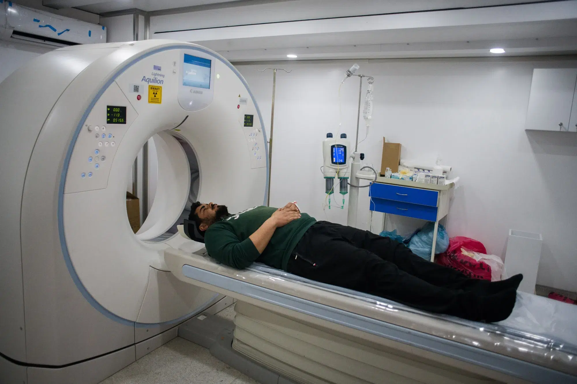
The scan experience is similar to a regular MRI. Patients lie on a table that slides into the MRI machine. The scan is painless and non invasive.
During the scan:
Clear instructions are given before the scan begins. Staff members monitor you throughout the procedure to ensure comfort and safety.
Diffusion weighted imaging uses MRI technology and does not involve radiation. This makes it a safe option for repeated imaging when medically required.
Safety considerations include:
Doctors recommend diffusion MRI only when the benefits outweigh any potential risks.
Doctors may suggest diffusion imaging when symptoms or test results require deeper evaluation.
Common reasons include:
It is always prescribed as part of a medical evaluation, not as a routine test without reason.
While diffusion imaging is powerful, it has limitations.
These include:
Doctors interpret diffusion findings alongside clinical history and other imaging results.
Radiologists analyze diffusion images carefully, comparing them with conventional MRI scans and clinical details.
Interpretation involves:
This collaborative approach ensures accurate diagnosis and appropriate care planning.
Diffusion weighted imaging is increasingly used as part of whole body MRI, especially in cancer screening, staging, and follow up. Unlike scans that focus on one organ, whole body diffusion MRI examines multiple regions in a single session. This helps doctors identify abnormal tissue activity across different organs at the same time.
Because diffusion imaging looks at water movement at a cellular level, it can highlight suspicious areas even before visible structural changes appear. This makes it useful for detecting early cancer spread, monitoring treatment response, and checking for disease recurrence. Since it does not use radiation, whole body diffusion MRI is suitable for repeated scans, long term monitoring, and patients who require ongoing evaluation.
Diffusion imaging is preferred in stroke cases because it detects brain changes much earlier than regular MRI. When a stroke occurs, blood flow to part of the brain is suddenly reduced. This causes brain cells to swell quickly, restricting water movement inside the tissue.
Diffusion weighted imaging captures this restriction within minutes to hours after stroke onset. Regular MRI sequences may appear normal during this early phase. Early detection is critical because treatment decisions depend on timing. Diffusion imaging helps doctors confirm stroke diagnosis, assess the extent of brain injury, and decide on urgent treatment options. This early clarity can significantly improve patient outcomes and reduce long-term complications.
Yes, diffusion weighted imaging can help detect cancer at an early stage. Cancer cells are usually tightly packed and abnormal in structure. This restricts the normal movement of water molecules within the tissue.
Diffusion imaging highlights these restricted areas, sometimes before tumors grow large enough to be seen clearly on other scans. This is especially helpful in identifying aggressive tumors, detecting spread, and monitoring response to treatment. While diffusion MRI alone does not confirm cancer, it provides valuable information that guides further testing and medical decisions. Early detection through diffusion imaging supports timely diagnosis and more effective treatment planning.
DWI is becoming an important tool for identifying cancer earlier and improving diagnostic accuracy. Explore Why Diffusion MRI Is Changing the Way Cancer Is Detected Early for a detailed overview.
In many cases, diffusion MRI does not require contrast injection. The technique relies on measuring water movement within tissues, which can be assessed without contrast agents. This is beneficial for patients who cannot receive contrast due to kidney issues or allergies.
However, in some situations, doctors may recommend contrast to gain additional information, especially when evaluating tumors or blood supply. The decision depends on the clinical question and the area being examined. The radiologist and referring doctor decide whether contrast is necessary. Patients are informed in advance, and safety checks are always performed before contrast administration.
A diffusion MRI scan usually takes between 20 and 45 minutes, depending on the body area being examined and whether it is part of a whole body MRI. The diffusion sequence itself is relatively quick, but it is often combined with other MRI sequences for a complete evaluation.
Patients are required to lie still during the scan to ensure clear images. The machine may produce loud sounds, but ear protection is provided. Most patients tolerate the scan well. After the scan, normal activities can usually be resumed immediately, unless advised otherwise by the medical team.
Restricted diffusion means water movement is limited, often due to cell swelling, infection, or dense tumor tissue.
No. The scan is painless and non invasive.
It is highly accurate, especially in early stroke diagnosis.
Yes. It helps distinguish between abscesses and tumors based on diffusion patterns.
Yes. It does not use radiation and is considered safe when medically advised.
No. It supports diagnosis but does not replace tissue biopsy when required.
Results are often available within one to two days, depending on the facility.
Diffusion Weighted Imaging contributes meaningful information by revealing early changes in tissue behavior. It plays a key role in stroke care, cancer evaluation, and infection assessment. Used alongside other tests and clinical evaluation, it supports more confident medical decisions. Discussing results with your doctor helps translate imaging findings into appropriate and timely care.
Whole body diffusion cancer screening offers a powerful way to detect abnormal cellular changes early and without radiation exposure. MVR Diagnostic Clinic provides trusted and advanced diagnostic care using high quality MRI technology and skilled medical professionals. With a strong focus on accurate results, patient comfort, and clear reporting, MVR supports informed medical decisions. Affordable screening and diagnostic packages are available, making advanced imaging accessible for individuals seeking reliable and timely cancer evaluation in Dubai.
WhatsApp us