

Persistent joint pain that continues for weeks can signal hidden damage inside the joint. When rest, medication, or basic tests do not explain the pain, doctors recommend MRI scans to see deeper structures clearly. MRI helps detect ligament tears, cartilage damage, inflammation, and muscle injuries. Early MRI diagnosis helps doctors plan proper treatment and prevent long term complications. Read on to know when MRI becomes necessary and how it supports recovery.
Joint pain that lasts longer than expected is often linked to internal joint problems. Unlike temporary pain caused by strain or overuse, persistent joint pain usually indicates structural damage, inflammation, or degeneration. Many patients ignore early symptoms, assuming the pain will resolve naturally, but delayed diagnosis can lead to worsening conditions.
Persistent joint pain can affect daily activities such as walking, climbing stairs, lifting objects, or even resting comfortably. When joint structures such as cartilage, ligaments, or tendons are damaged, the body cannot repair them properly without medical support. MRI scans help doctors identify the exact cause of pain and recommend appropriate treatment.
Common causes of persistent joint pain include:
Early imaging helps prevent further joint damage and improves recovery outcomes.
Doctors do not recommend an MRI immediately for all joint pain. Initial evaluation usually includes physical examination, medical history review, and basic imaging such as X-ray. However, when pain continues without a clear explanation, an MRI becomes necessary.
MRI scans provide detailed images of soft tissues, which are not visible in X-ray scans. This helps orthopaedic specialists confirm whether the pain is caused by ligament tears, cartilage damage, or muscle injuries.
Doctors commonly recommend an MRI in the following situations:
MRI helps doctors avoid incorrect treatments and ensures accurate diagnosis.
MRI scan is considered the most advanced imaging technique for evaluating joint and muscle problems. Unlike X-ray, which mainly shows bones, MRI provides clear images of soft tissues such as ligaments, cartilage, tendons, and muscles.
This detailed imaging allows doctors to detect problems at early stages before severe damage develops. MRI is especially useful when pain is caused by internal injuries that are not visible through basic imaging.
MRI scan can detect the following conditions:
MRI provides precise imaging that helps doctors plan correct treatment.
To better understand how MRI compares with other imaging techniques, read our detailed guide on Difference Between MRI and CT Scan.
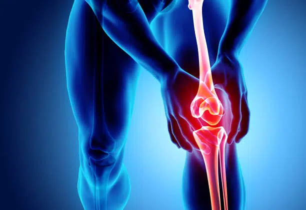
Knee pain is one of the most common reasons doctors recommend MRI scan. The knee joint contains complex structures such as ligaments, cartilage, and tendons that cannot be fully evaluated with X ray.
MRI helps detect internal knee injuries that cause persistent pain and mobility problems. Early diagnosis allows doctors to recommend proper treatment and prevent long term joint damage.
MRI scan helps identify:
MRI is essential when knee pain continues after injury or treatment.
Learn what to expect from the procedure in our guide, How Long Does a Knee MRI Take?.
Shoulder pain often develops due to muscle strain, tendon injury, or ligament damage. Since the shoulder joint allows wide range of motion, it is vulnerable to injuries from sports, lifting, and repetitive activities.
MRI scan provides clear images of shoulder structures, helping doctors diagnose the exact cause of pain. This allows targeted treatment that improves recovery and restores mobility.
MRI can detect:
MRI helps prevent worsening of shoulder injuries.
Hip pain can develop due to cartilage damage, muscle injury, or joint degeneration. Many hip problems are not visible on X ray, especially in early stages.
MRI scan helps doctors identify hidden causes of hip pain and recommend appropriate treatment.
MRI helps detect:
MRI supports early diagnosis and faster recovery.
Persistent joint pain combined with specific symptoms often indicates internal joint damage. These symptoms should not be ignored, as delayed diagnosis may worsen the condition.
Common warning signs include:
MRI scan helps identify the cause and guides treatment planning.
Arthritis is a common cause of persistent joint pain, especially in older adults. Early diagnosis helps slow disease progression and prevent severe joint damage.
MRI scan can detect arthritis changes before they appear on X ray. This allows doctors to begin treatment early and reduce joint damage.
MRI helps identify the following arthritis related changes:
Early MRI diagnosis improves treatment effectiveness.
MRI scan is a safe and painless procedure used to evaluate joint and muscle problems. It does not use radiation and is suitable for repeated diagnostic use when necessary.
During the MRI scan procedure, the patient lies on a scanning table that moves into the MRI machine. The machine uses magnetic fields to create detailed images of the joint.
MRI scan procedure includes:
MRI provides clear images that help doctors diagnose joint problems.
MRI scan cost in UAE varies depending on the joint area, diagnostic center, and whether contrast is used. Advanced imaging technology and radiologist expertise also influence cost.
Typical MRI scan costs in UAE include:
MRI scan is one of the safest imaging methods available. It does not use harmful radiation and is suitable for diagnosing joint and muscle conditions.
MRI scan safety advantages include:
MRI is widely used for orthopedic diagnosis.
MRI and X ray serve different diagnostic purposes. X ray is useful for detecting bone fractures, while MRI provides detailed images of soft tissues.
MRI is more effective for diagnosing ligament injuries, cartilage damage, and muscle problems. It helps doctors identify conditions that X ray cannot detect.
MRI is preferred when joint pain persists without clear diagnosis. It provides accurate imaging and supports proper treatment planning.
MRI scan provides detailed information that helps doctors recommend effective treatment. Accurate diagnosis reduces treatment delays and improves recovery outcomes.
MRI helps doctors in the following ways:
MRI improves treatment accuracy and patient recovery.
Joint pain that lasts longer than normal recovery time requires medical evaluation. Early diagnosis helps prevent serious joint damage and improves treatment success.
You should consult a doctor if you experience the following:
Early MRI scan helps diagnose joint problems accurately.
Persistent joint pain should never be ignored, especially when symptoms continue beyond normal recovery time. MRI scan provides detailed imaging that helps detect ligament injuries, cartilage damage, arthritis, and muscle problems. Accurate diagnosis allows doctors to recommend effective treatment and prevent further joint damage. MRI plays a critical role in orthopedic diagnosis and helps patients recover safely and return to normal activities with improved joint health.
MVR provides trusted and advanced MRI scan services in Dubai using high quality imaging technology and experienced medical professionals. Our diagnostic approach focuses on accurate results, patient comfort, and reliable evaluation of joint and muscle conditions. We offer affordable MRI scan packages designed to support early diagnosis and treatment planning. Patients benefit from professional care, modern facilities, and precise diagnostic imaging for confident medical decisions and faster recovery.
If joint pain lasts more than six weeks, worsens with movement, or shows swelling and stiffness, doctors recommend MRI to detect ligament injuries, cartilage damage, or inflammation accurately.
Yes, MRI is the best imaging method to detect ligament tears, tendon injuries, cartilage damage, and muscle problems. It provides clear images that help doctors confirm the cause of joint pain.
MRI is better for detecting soft tissue injuries, cartilage damage, and ligament tears. X ray only shows bones, while MRI provides detailed imaging of internal joint structures.
MRI scan usually takes between twenty and forty-five minutes. The exact time depends on the joint being scanned and whether contrast imaging is required.
MRI is very safe because it does not use radiation. It uses magnetic fields to create detailed images and is suitable for diagnosing joint and muscle conditions.
MRI scan cost in Dubai ranges from AED 900 to AED 3500 depending on the joint, contrast use, and diagnostic center technology and expertise.
WhatsApp us