

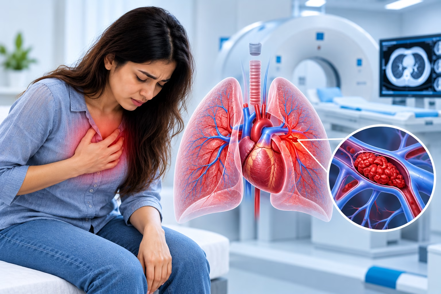
A pulmonary embolism (PE) is a blockage in one of the arteries in the lungs, usually caused by a blood clot that has traveled from elsewhere in the body, most often the legs. It is a serious and potentially life-threatening condition that requires prompt diagnosis and treatment.
If you are experiencing sudden breathlessness, chest pain, or other symptoms of a possible pulmonary embolism, seek emergency medical care immediately. CT pulmonary angiography can help doctors identify blood clots in the lung arteries. This guide explains pulmonary embolism symptoms, diagnosis, imaging, and treatment options.
Sudden severe breathlessness, chest pain, fainting, or coughing up blood can indicate a pulmonary embolism. Seek emergency medical attention rather than waiting for a scan appointment.
A pulmonary embolism, often abbreviated as PE, occurs when a blood clot blocks one or more arteries supplying blood to the lungs. Most clots originate in the deep veins of the legs or pelvis, a condition known as deep vein thrombosis (DVT). The clot can travel through the bloodstream and become lodged in the lungs.
Depending on the size and number of clots, a pulmonary embolism can range from relatively mild to life-threatening. A large clot can significantly block blood flow through the lungs, reduce oxygen levels, and place strain on the heart.
A pulmonary embolism is most commonly caused by a blood clot that has traveled from another part of the body, particularly the deep veins of the legs.
Long periods of sitting during flights or car journeys, or extended bed rest after surgery or illness, can slow blood flow in the legs and increase clotting risk.
Major surgery, particularly orthopedic procedures, and significant injuries can increase the risk of blood clots during recovery.
Certain cancers and cancer treatments can increase the tendency of blood to clot.
Pregnancy changes blood clotting factors and can increase pressure on pelvic veins. The risk remains elevated in the weeks after delivery.
Estrogen-containing birth control pills and hormone replacement therapy can increase clotting risk in some people.
Inherited clotting disorders, excess body weight, and smoking can contribute to an increased risk of blood clots.
Factors associated with a higher risk of pulmonary embolism include:
A pulmonary embolism can also occur in people without obvious risk factors. The absence of known risk factors does not rule out the condition.
Symptoms can vary depending on the size of the clot and how much of the lung's blood supply is affected. Common symptoms include:
Symptoms can sometimes be subtle, particularly with smaller clots. If you have ongoing breathlessness or a cough, you can read the guide on CT scanning and lung evaluation for general information about chest imaging. However, suspected pulmonary embolism requires urgent medical assessment.
Large pulmonary emboli can strain the heart and reduce oxygen levels throughout the body. Without prompt treatment, the condition can be life-threatening.
Diagnosis usually begins with a review of symptoms, medical history, and risk factors, followed by a physical examination. Because symptoms can overlap with other conditions, including a heart attack or pneumonia, doctors may use a combination of clinical assessment and tests.
A D-dimer blood test measures a substance released when blood clots break down. In people considered to be at lower risk, a normal D-dimer result can help doctors rule out pulmonary embolism.
An elevated D-dimer result does not confirm a pulmonary embolism by itself because many other conditions can increase the level. Depending on the clinical situation, additional blood tests may be used to assess overall health and possible heart strain.
Blood test results must be interpreted alongside symptoms, examination findings, and other investigations. Your doctor will determine which tests are appropriate.
CT pulmonary angiography, commonly called CTPA, is a specialized CT scan used to investigate suspected pulmonary embolism. It uses an intravenous contrast injection to highlight the blood vessels in the lungs, allowing clots within the pulmonary arteries to be visualized.
CT pulmonary angiography provides detailed images of the lung arteries and can help doctors identify the location and extent of a clot. Rapid imaging can be particularly important when pulmonary embolism is suspected because the condition may require urgent treatment.
For a broader explanation of CT imaging, read the CT scan guide.
On CT pulmonary angiography, a pulmonary embolism typically appears as a filling defect, where contrast material does not pass normally through a blood vessel because a clot is blocking it.
The scan can help show:
CT pulmonary angiography is widely used as a first-line imaging test when pulmonary embolism is suspected and is highly accurate when performed and interpreted appropriately.
You will usually lie on your back on the CT table. A contrast agent is injected into a vein, typically in your arm, and images are acquired as the contrast passes through the blood vessels in your lungs.
You may be asked to hold your breath briefly to reduce motion blur. Image acquisition usually takes only a few minutes, although preparation, positioning, and the contrast injection may take additional time.
The imaging team reviews relevant medical history, contrast-related concerns, and any preparation instructions.
You remain still on the CT table while images are captured during the contrast injection. Follow the team's breathing instructions.
The images are reviewed by a radiologist, and the findings are communicated to the treating medical team.
Before the scan, tell your doctor or imaging team if you:
Because the test uses contrast material, you may be asked to fast for a short period. Kidney function may also be checked before the scan, particularly if you have existing kidney concerns. Follow the specific instructions provided by your treating doctor and imaging team.
CT pulmonary angiography involves exposure to a small amount of ionizing radiation and the use of intravenous contrast material. Contrast can cause an allergic reaction in some people and may affect kidney function in certain patients, particularly those with existing kidney problems.
Your doctor will consider your medical history and individual circumstances when assessing the risks and benefits. When pulmonary embolism is suspected, timely and accurate diagnosis is important because an untreated clot can be dangerous.
Depending on your symptoms and clinical situation, your doctor may recommend other tests alongside or instead of CT pulmonary angiography.
Checks for deep vein thrombosis, which is a common source of the clot causing pulmonary embolism.
Uses ultrasound to assess the heart and may help evaluate strain in more significant cases.
May be used to assess other lung conditions, although a routine chest CT is not the same as CT pulmonary angiography.
A V/Q scan evaluates air and blood flow in the lungs and may be considered in selected situations, including when CT contrast cannot be used.
Treatment depends on the clot's size, the person's overall health, and whether the heart is under strain. A treating medical team determines the most appropriate approach.
Blood-thinning medication is the main treatment for most pulmonary embolisms. These medicines help prevent new clots from forming while the body's natural processes gradually break down the existing clot.
Clot-dissolving medication may be used for large or life-threatening pulmonary emboli. Because it carries a higher risk of bleeding, it is generally reserved for selected severe cases.
In selected severe cases, a procedure may be used to remove or break down the clot, particularly when medication alone is not appropriate or effective.
Oxygen therapy and other supportive measures may be required to manage symptoms and support the patient, especially in more serious cases.
The duration of blood-thinning treatment varies depending on the cause of the clot and whether risk factors remain. Your doctor will determine the appropriate treatment duration and follow-up plan.
Certain measures can help reduce the risk of blood clots, particularly for people with known risk factors.
People with a previous DVT or pulmonary embolism should follow their clinician's advice about prevention, medication, and follow-up care.
Seek prompt medical assessment if you experience:
Because pulmonary embolism can be life-threatening, do not wait for symptoms to improve on their own. If symptoms are severe or sudden, seek emergency medical care immediately.
MVR Diagnostic Clinic in Al Garhoud, Dubai, offers CT imaging using advanced low-dose, AI-enhanced CT technology. According to the clinic's stated service information, reports are reviewed by DHA-licensed consultant radiologists and delivered digitally within 24 hours.
If your doctor has recommended CT pulmonary angiography, contact the clinic to discuss the examination and appointment arrangements. If you are experiencing symptoms of a possible pulmonary embolism, however, seek emergency care first rather than waiting for a scheduled outpatient scan.
Contact MVR Diagnostic Clinic in Al Garhoud, Dubai, for information about CT imaging appointments and preparation requirements.
Contact MVR Diagnostic Clinic Enquire on WhatsAppLearn more about available CT scan services in Dubai.
Common warning signs include sudden breathlessness, chest pain that worsens with breathing, a rapid heartbeat, coughing that may include blood, and lightheadedness. These symptoms require prompt medical evaluation, and severe or sudden symptoms warrant emergency care.
CT pulmonary angiography is a specialized CT scan used to visualize blood clots in the pulmonary arteries and help confirm or rule out a suspected pulmonary embolism.
The scan itself is generally painless. You may experience a brief warm sensation when the contrast material is injected. You will need to remain still and may be asked to hold your breath briefly during imaging.
CT pulmonary angiography usually takes only a few minutes to acquire images. The time needed for preparation, interpretation, and communication of results can vary. Suspected pulmonary embolism requires prompt medical assessment.
Yes. Many pulmonary embolisms are treated with anticoagulant medication. Clot-dissolving medicines or catheter-based and surgical procedures may be considered for selected severe cases.
Yes. Recurrence is possible, particularly if underlying risk factors remain. Your doctor will advise on the appropriate treatment duration, follow-up, and preventive measures based on your individual risk.
Medical disclaimer: This article is for general educational purposes and does not replace professional medical advice, diagnosis, or treatment. A suspected pulmonary embolism is a medical emergency. Seek immediate medical attention for sudden severe breathlessness, chest pain, fainting, or coughing up blood.
WhatsApp us