

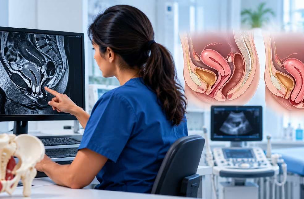
Uterine prolapse occurs when the muscles and ligaments supporting the uterus weaken, causing the uterus to descend into or, in more severe cases, out of the vaginal canal. It is a common condition, particularly among women who have had vaginal childbirth or who are past menopause. Symptoms can range from mild and barely noticeable to significantly affecting daily life.
If you are experiencing pelvic pressure, a bulge, or discomfort that may suggest prolapse, a medical evaluation can help identify the cause. This guide explains uterine prolapse symptoms, the role of pelvic imaging, diagnosis, and treatment options.
Looking for pelvic imaging in Dubai? Contact MVR Diagnostic Clinic to enquire about pelvic ultrasound appointments.
The uterus is normally held in place within the pelvis by a network of muscles and ligaments collectively known as the pelvic floor. When these supporting structures weaken or stretch, the uterus can shift downward from its normal position. This condition is called uterine prolapse.
Uterine prolapse is commonly described in stages according to how far the uterus has descended:
The uterus has dropped slightly but remains well within the vagina.
The uterus has descended closer to the vaginal opening.
Part of the uterus protrudes outside the vaginal opening.
The uterus is entirely outside the vaginal opening. This is also called procidentia.
Uterine prolapse can occur alongside prolapse of other pelvic organs, such as the bladder or rectum, because these organs share many of the same supporting structures.
Uterine prolapse develops when the pelvic floor muscles and ligaments become weakened or damaged over time. Several factors may contribute to this process.
Vaginal delivery, particularly multiple deliveries, prolonged labor, or delivery of a larger baby, can stretch and weaken the pelvic floor muscles and supporting ligaments.
Declining estrogen levels after menopause can reduce the strength and elasticity of pelvic tissues, making prolapse more likely to develop or worsen.
The natural aging process can gradually weaken pelvic floor support, regardless of other contributing factors.
Chronic coughing, long-term constipation with straining, and repeated heavy lifting can place ongoing pressure on the pelvic floor.
Excess body weight adds pressure to the pelvic organs and their supporting tissues.
Some women naturally have weaker connective tissue, which can make them more prone to prolapse even without other risk factors.
Certain previous pelvic surgeries can affect the structures that support the uterus.
Factors that may increase the likelihood of developing uterine prolapse include:
However, uterine prolapse can also occur in women who do not have these risk factors.
Symptoms vary according to the severity of the prolapse. Mild cases may cause no noticeable symptoms, while more significant prolapse can affect everyday comfort and function.
Common symptoms include:
Symptoms often become more noticeable toward the end of the day or after prolonged standing and may improve after lying down.
Yes. As the uterus descends, it can alter the normal anatomy of the vagina, potentially causing discomfort, pain, or a feeling of pressure during intercourse. This symptom may prompt women to seek medical evaluation and discuss treatment options with their healthcare provider.
Pain during intercourse can have several causes, so it is important not to assume that prolapse is responsible without an appropriate examination.
Yes. The bladder and rectum are closely connected to the same supporting structures as the uterus. Prolapse can therefore affect their function in some women.
When these organs are also affected, it may indicate a combined prolapse involving more than one pelvic structure. A healthcare professional can assess the symptoms and determine whether further testing is needed.
Diagnosis usually begins with a discussion of symptoms and medical history, including childbirth history, followed by a physical examination. Your doctor may ask you to bear down or cough during the examination to assess the degree of prolapse.
Common diagnostic steps include:
A physical examination is often sufficient to diagnose uterine prolapse. However, pelvic ultrasound can provide additional information, particularly when the uterus, bladder, and surrounding structures need to be assessed together. A transvaginal ultrasound may also be used to obtain more detailed images of the pelvic organs.
Explore MVR Diagnostic Clinic's ultrasound services to learn more about available examinations.
Pelvic ultrasound may show a lower-than-normal position of the uterus within the pelvis. Imaging performed while the patient strains or bears down may help demonstrate the degree of descent in appropriate cases.
Ultrasound can also help assess whether the bladder or rectum may be involved and identify other conditions that could contribute to pelvic symptoms.
For example, uterine fibroids can cause pelvic pressure or discomfort. Read more in our guide to uterine fibroids, their causes, symptoms, and ultrasound findings.
Imaging findings need to be interpreted alongside symptoms and physical examination results. Ultrasound is not a replacement for a pelvic examination when assessing suspected uterine prolapse.
The pelvic floor supports multiple organs, so uterine prolapse can occur alongside other forms of pelvic organ prolapse.
The bladder bulges into the front wall of the vagina. This may contribute to urinary symptoms, including difficulty emptying the bladder or urinary leakage.
The rectum bulges into the back wall of the vagina, which can contribute to difficulty with bowel movements.
In women who have had a hysterectomy, the top of the vagina can descend because of weakened supporting structures.
Identifying whether more than one pelvic structure is involved helps the healthcare team plan an appropriate treatment approach.
Treatment depends on the severity of the prolapse, the symptoms, age, general health, and whether the patient wishes to preserve fertility. Not every case requires surgery.
Also known as Kegel exercises, these exercises strengthen the pelvic floor muscles and may improve symptoms in mild to moderate prolapse, particularly when started early. A healthcare professional or pelvic floor physiotherapist can help ensure the exercises are performed correctly.
Managing chronic cough, treating constipation, maintaining a healthy body weight, and avoiding excessive heavy lifting can help reduce strain on the pelvic floor and may slow progression.
A vaginal pessary is a removable device placed inside the vagina to support the uterus and surrounding structures. It is a nonsurgical option that may be suitable for women who prefer to avoid or delay surgery.
For some postmenopausal women, topical estrogen may be recommended to help improve the condition of vaginal tissues. It may be used alongside other treatments when appropriate.
Surgery may be considered for more significant prolapse or when other treatments do not provide sufficient relief. Options range from procedures that preserve the uterus to those that remove it, depending on the individual's circumstances, the severity of the prolapse, and future fertility plans.
Important: Treatment should be individualized. A gynecologist or urogynecology specialist can explain the benefits and limitations of available options based on your symptoms and examination findings.
Not all cases of uterine prolapse can be prevented, but certain measures may help reduce the risk or slow progression:
These measures may help reduce strain on the pelvic floor, but they cannot guarantee that prolapse will not develop.
Arrange a medical evaluation if you experience:
Early evaluation can help confirm the diagnosis and guide appropriate treatment. Seek prompt medical advice if you have difficulty emptying your bladder, significant pain, or new or unexplained vaginal bleeding.
MVR Diagnostic Clinic in Al Garhoud, Dubai, offers pelvic and transvaginal ultrasound examinations. According to the clinic's stated service information, reports are reviewed by DHA-licensed consultant radiologists and delivered digitally within 24 hours.
If your doctor has recommended pelvic imaging to investigate your symptoms, contact the clinic to enquire about available appointments and the examination that may be appropriate for you.
Contact MVR Diagnostic Clinic in Dubai to enquire about pelvic and transvaginal ultrasound appointments.
Contact MVR Diagnostic Clinic Enquire on WhatsAppLearn more about available ultrasound services.
Early signs can include pelvic pressure or heaviness, mild lower back pain, and a sensation that something is bulging in the vagina, particularly after standing for long periods. Some women with mild prolapse have no noticeable symptoms.
Yes. Pelvic ultrasound may help demonstrate the position and descent of the uterus. Imaging performed with straining may provide additional information in appropriate cases. A pelvic examination is also important for assessing prolapse.
No. Many cases, particularly mild to moderate prolapse, can be managed with pelvic floor exercises, lifestyle changes, or a vaginal pessary. Surgery may be considered for more significant prolapse or when other treatments are not effective.
Yes. Prolapse can progress over time, although the course varies between individuals. Early evaluation and management of contributing factors may help manage symptoms and reduce further strain on the pelvic floor.
Yes. Vaginal childbirth, especially multiple deliveries, is an important risk factor because delivery can stretch and weaken the pelvic floor muscles and supporting ligaments.
Pelvic floor exercises can strengthen supporting muscles and improve symptoms, particularly in mild cases, but they may not fully reverse more advanced prolapse. A healthcare professional can advise on suitable treatment options.
Medical disclaimer: This article is for general educational purposes and is not a substitute for professional medical advice, diagnosis, or treatment. Consult a qualified healthcare professional for assessment of suspected uterine prolapse and personalized treatment recommendations.
WhatsApp us