
If you’ve been coughing without relief, feeling short of breath, or an X-ray shows something odd, then a chest CT scan might be the next step. A CT or CAT scan on lungs provides clearer, more detailed cross-sectional images than a regular chest X-ray. This guide explains when you might need a CT chest scan, what lung problems it can detect, and how it helps your doctor make faster and more accurate decisions.
What is a CT chest scan and how it differs from a regular chest X-ray
A CT chest scan, also called a chest computed tomography scan or CAT scan on lungs, uses X-ray technology with computer processing to create detailed cross-sectional images of your chest. These images show the lungs, blood vessels, airways, and chest cavity in far greater detail than a normal X-ray.
While a standard chest X-ray gives a flat, two-dimensional image, a CT chest scan provides 3D views, allowing doctors to see even tiny nodules, infections, or blockages that an X-ray may miss. This makes it especially useful for diagnosing complex or hidden lung problems.
Differences between CT and X-ray:
- CT scans use rotating X-ray beams to capture multiple angles.
- Images are reconstructed into detailed cross-sections.
- They reveal small or early changes in lung tissue.
- A CT scan can differentiate between fluid, tissue, and air.
Some patients may also be recommended a low-dose lung CT scan, which uses less radiation and is often used for early lung cancer screening in long-term smokers or people with high risk factors.
When should you consider a CT scan of your lungs?
A CT chest scan is not something done routinely. It is ordered when there are signs that something deeper may be going on in the lungs.
You might be asked to undergo a lung CT scan if you:
- Have a persistent cough that doesn’t improve after treatment.
- Experience chest pain, tightness, or difficulty breathing.
- Have abnormal findings on a chest X-ray.
- Are a smoker or ex-smoker with risk factors for lung disease.
- Have a known lung condition that needs close monitoring.
Doctors recommend this scan for several reasons:
- To detect infections or tumors not visible on X-rays.
- To assess the spread of a disease.
- To monitor how well a treatment is working.
- To guide procedures like biopsies or surgeries.
Typical places where CT chest scans are done:
- Hospitals and diagnostic imaging centers.
- Specialized radiology departments.
- Clinics with advanced imaging systems.
A referral is usually given by your doctor after evaluating your symptoms or test reports. In screening programs, especially for lung cancer, low-dose CT scans may be offered yearly to people at high risk.
Common lung problems that a chest CT scan can detect
A chest CT scan is one of the most powerful diagnostic tools in respiratory medicine. It helps uncover several lung problems that may not show up clearly in a basic X-ray. Below are some of the most common ones.
Lung nodules and early lung cancer
Lung nodules are small, round spots that can appear in the lung tissue. Most are harmless, often caused by healed infections or inflammations, but some may be early signs of lung cancer.
A CT chest scan can detect even very small nodules that X-rays often miss. This allows doctors to:
- Track the size and shape of nodules over time.
- Compare current and previous scans for changes.
- Identify signs that may suggest early cancer.
For high-risk individuals, low-dose CT lung screening is recommended annually to detect cancer before symptoms appear. Early detection often means simpler and more effective treatment.
Pulmonary embolism (blood clot in lung)
A pulmonary embolism occurs when a blood clot travels to the lungs and blocks a blood vessel. It can cause sudden chest pain, shortness of breath, and rapid heartbeat, and it requires immediate medical attention.
A specialized form of chest CT called CT pulmonary angiography helps visualize the blood vessels and detect blockages. It is considered the gold standard for diagnosing this condition.
Typical symptoms prompting this scan include:
- Sharp chest pain that worsens with breathing.
- Unexplained breathlessness.
- Coughing up small amounts of blood.
Pneumonia, infections and complications
While pneumonia is often diagnosed using a chest X-ray, sometimes a CT scan is needed for a clearer picture. This helps confirm the extent of infection, detect abscesses, or rule out other conditions.
A lung CT scan is especially useful in:
- Severe or long-lasting pneumonia.
- Recurrent infections not responding to antibiotics.
- Post-COVID lung changes.
It helps doctors determine how much of the lung is affected and whether there is fluid, scarring, or other complications.
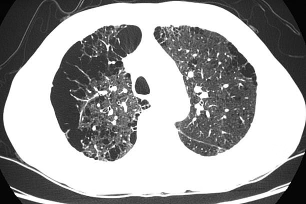
Interstitial lung disease and fibrosis
Interstitial lung disease (ILD) is a group of conditions that cause inflammation and scarring of lung tissue. A high-resolution CT scan (HRCT) is the best way to detect and monitor this condition.
The scan can reveal:
- Patterns of fibrosis (thickened tissue).
- Honeycombing or scarring in the lungs.
- How much lung capacity is lost over time.
ILDs often develop slowly and can be missed on simple X-rays. Regular CT monitoring helps doctors plan early treatment to slow down disease progression.
Pleural disorders and chest trauma
The pleura are thin layers surrounding the lungs. CT scans help identify pleural effusion (fluid buildup) or pneumothorax (air leakage).
Chest trauma from accidents or falls may also require a CT chest scan to assess internal injuries. It helps detect:
- Rib fractures.
- Bleeding inside the chest cavity.
- Air or fluid compressing the lungs.
A detailed CT image ensures no hidden injury is missed, making it crucial in emergency and post-trauma assessments.
What to expect during and after your lung CT scan
A CT chest scan is quick, non-invasive, and generally takes less than 10 minutes. Knowing what to expect helps reduce anxiety and ensures smooth preparation.
Before the scan:
- You may be asked to avoid eating for a few hours if contrast dye is used.
- Remove metal items like jewelry, belts, or watches.
- Wear comfortable clothing or a hospital gown.
During the scan:
- You will lie on a moving table that slides into the scanner.
- The machine rotates around your chest while taking images.
- You may be asked to hold your breath for a few seconds to avoid blurring.
After the scan:
- You can return to normal activities right away.
- If contrast dye was used, drink water to help flush it out.
- Results are usually available within the same day or shortly after.
Safety note:
Radiation exposure from a CT scan is minimal and considered safe for adults. For screenings, low-dose protocols are used to reduce risk further.
Risks, benefits and how to choose the right time for a scan
Like any medical procedure, a CT chest scan has both benefits and minor risks. Understanding them helps you make informed choices.
Benefits of a CT chest scan:
- Provides highly detailed images for accurate diagnosis.
- Detects small changes early, allowing faster treatment.
- Helps doctors plan surgeries, biopsies, or therapies.
- Monitors ongoing lung disease effectively.
- Plays a key role in preventive health for high-risk patients.
Possible risks:
- Slight radiation exposure.
- Mild allergic reaction to contrast dye (rare).
- False positives that may lead to further tests.
You should discuss your medical history, smoking habits, and symptoms with your doctor to determine whether a low-dose screening CT or a full diagnostic CT is needed. Screening scans are ideal for long-term smokers or people with family history of lung cancer, while full scans are done to diagnose or track known lung problems.
Choosing the right timing helps avoid unnecessary exposure while still ensuring early detection when it matters most.
FAQs – Answers to common questions
- What is the difference between a chest CT scan and a chest X-ray?
A chest X-ray shows a broad image of the lungs and heart, while a CT scan provides detailed 3D cross-sections that can reveal smaller abnormalities like nodules or infections. - When does a doctor order a CT scan of the lungs instead of relying on an X-ray?
A CT scan is ordered when symptoms or X-ray results suggest something more complex, such as unexplained cough, chest pain, or signs of a tumor or clot. - Does everyone with a cough need a CT chest scan on lungs?
No. Most coughs are caused by infections or allergies. A CT scan is suggested only if the cough is persistent or linked to other warning signs like blood in sputum or weight loss. - What does a low-dose CT scan for lung screening involve?
It’s a quick, painless scan that uses minimal radiation to screen for early lung cancer in people with high-risk factors, especially long-term smokers. - Are CT scans safe for lung screening? What about radiation risk?
Modern CT machines use very low doses of radiation. The benefits of early detection far outweigh the small risk, especially in high-risk groups. - How quickly will I get results from a CT chest scan and what happens next?
Reports are usually ready the same day or within 24 hours. Your doctor will explain the findings and recommend next steps such as medication, follow-up imaging, or specialist referral. - If my CT scan shows a lung nodule, does it mean I have lung cancer?
Not necessarily. Most nodules are benign. Your doctor may suggest periodic follow-up scans to monitor for any changes in size or shape. - How often should I get a CT chest scan if I am a former smoker?
Annual low-dose CT scans are recommended for adults aged 50–80 with a heavy smoking history, even if they quit within the last 15 years. - What should I do to prepare for a CT lung scan?
Avoid wearing metal objects, follow fasting instructions if contrast is used, and inform your doctor of any allergies or kidney issues. - Can children or pregnant women have a CT chest scan for lung problems?
CT scans are generally avoided during pregnancy unless absolutely necessary. For children, they are done only when the benefits clearly outweigh the risks.
A clear view for stronger lungs
A CT chest scan gives doctors the clarity they need to detect lung problems early and treat them effectively. From simple infections to serious conditions like lung cancer or pulmonary embolism, this advanced imaging test helps uncover what cannot be seen through standard X-rays. Whether it’s a CAT scan on lungs or a low-dose screening CT, the right test at the right time can make all the difference in protecting your respiratory health.
Advanced and Affordable Chest CT Scans at MVR
Experience precise imaging and expert care with MVR’s advanced chest CT scans. Our modern equipment and skilled radiologists ensure accurate results for lung and chest evaluations at affordable prices. Book your scan today for quick, reliable, and high-quality diagnostics you can trust.