

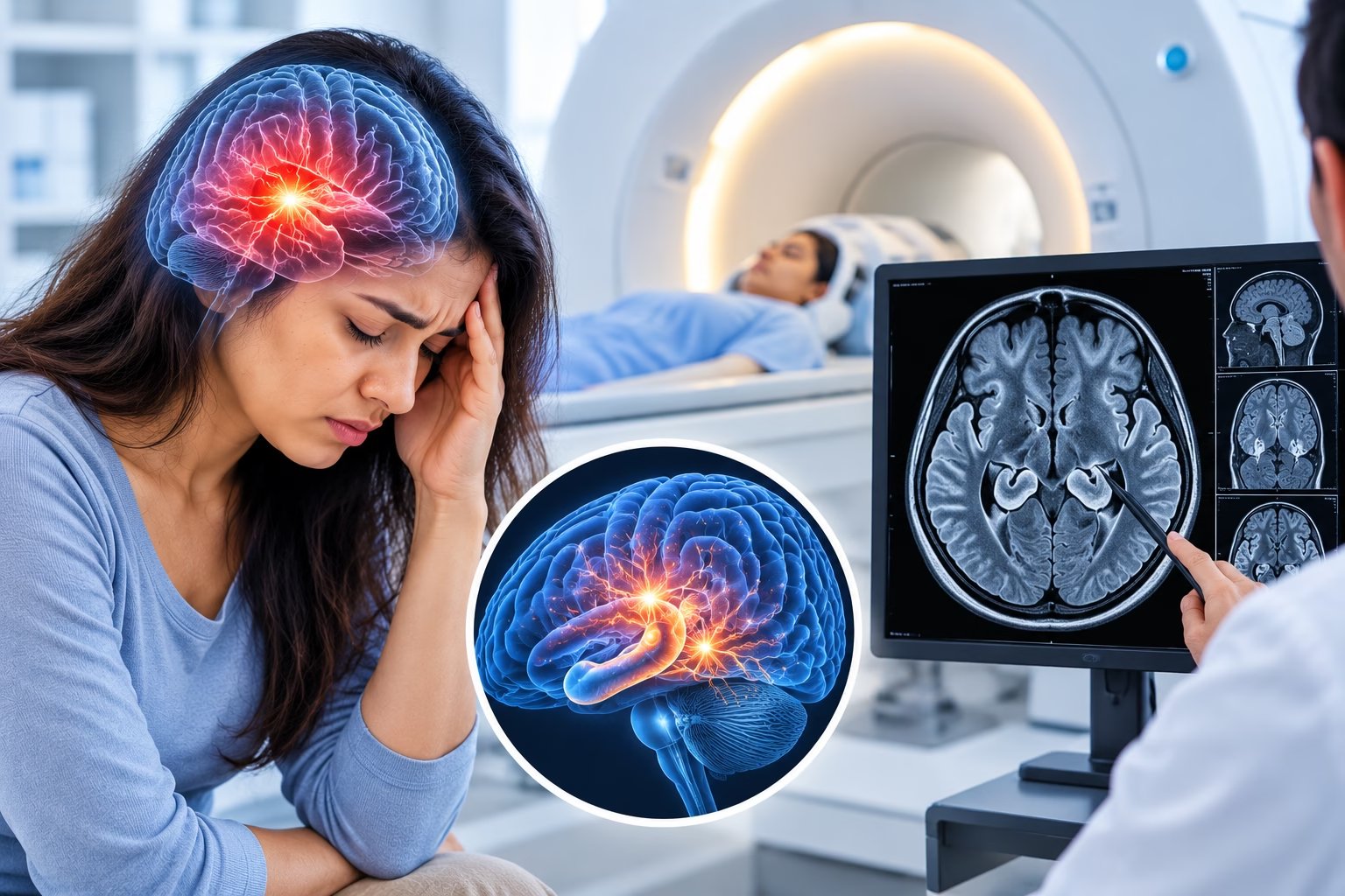
A seizure can be an alarming experience, whether it happens for the first time or as part of an ongoing condition. A brain MRI is one of the key imaging tests used to investigate seizures, helping doctors look for structural causes within the brain that may be contributing to abnormal electrical activity.
If you or a loved one has experienced a seizure, a brain MRI may help doctors investigate the underlying cause. This guide explains what a brain MRI can reveal about seizures, how the procedure works, and what the diagnostic process involves.
Looking for a brain MRI in Dubai? Contact MVR Diagnostic Clinic to discuss your scan and appointment requirements.
When someone experiences a seizure, doctors need to determine whether it was triggered by a temporary factor, such as fever, low blood sugar, or lack of sleep, or whether it points to an underlying structural or neurological condition.
Brain MRI is one of the most useful imaging tests for this purpose because it provides detailed images of the brain's structure. It can help identify abnormalities that may be causing or contributing to seizures.
MRI does not replace a clinical evaluation or an electroencephalogram (EEG). These tests provide different types of information:
Doctors may use both tests alongside a medical history and neurological examination to understand the cause of a seizure.
A brain MRI can help identify a range of structural abnormalities associated with seizures. The findings depend on the individual's symptoms, medical history, and the type of MRI performed.
Scarring or shrinkage of the hippocampus, a brain structure involved in memory, is a common finding associated with temporal lobe epilepsy.
MRI can detect noncancerous or cancerous tumors that may affect surrounding brain tissue and contribute to seizures.
Abnormal formations of blood vessels, including arteriovenous malformations and cavernomas, can sometimes be associated with seizure activity.
Some people are born with areas of the brain that did not develop typically. Cortical dysplasia can cause seizures, particularly in children and young adults.
Scar tissue following a stroke, head injury, or brain infection may act as a focus for abnormal electrical activity.
Past or current infections affecting the brain, such as encephalitis, may sometimes produce MRI findings associated with seizures.
Many people who experience seizures, particularly after a first seizure, have a normal brain MRI. This does not rule out epilepsy. Some causes of seizures are related to the brain's electrical function and may not produce visible structural changes on an MRI scan.
Your doctor may recommend a brain MRI if you have experienced any of the following:
In some children with certain well-understood seizure types that are unlikely to have a structural cause, MRI may not always be the first test recommended. The decision depends on the child's clinical presentation and the doctor's assessment.
Diagnosis usually begins with a detailed description of what happened before, during, and after the seizure. Information from someone who witnessed the episode can be particularly helpful. The doctor will also review the person's medical history and perform a neurological examination.
Common diagnostic steps include:
The tests required vary according to the person's age, symptoms, medical history, and the circumstances of the seizure.
During the examination, you will usually lie on your back on the MRI table, with your head positioned inside a dedicated head coil. The table moves into the scanner while images are captured using sequences designed to examine brain structures associated with seizures.
The scanner produces loud knocking or tapping sounds, so hearing protection is generally provided. Remaining still during the examination is important because movement can affect image quality.
A brain MRI does not require surgery and uses magnetic fields and radio waves to produce detailed images.
MRI does not use ionizing radiation, unlike X-ray-based imaging methods such as CT scans.
For details about MRI examinations available at MVR Diagnostic Clinic, visit the MRI scan services page.
A standard brain MRI typically takes around 30 to 45 minutes. A dedicated seizure-protocol MRI, which uses specific sequences to examine areas commonly involved in epilepsy, may take somewhat longer.
The expected duration depends on the imaging protocol and whether additional sequences or contrast images are needed. Your imaging team can explain the expected scan time before the examination begins.
Before your appointment: Ask the imaging team how long your specific scan is expected to take and whether you need to follow any preparation instructions.
Preparation depends on whether contrast is required and on the specific imaging protocol. Before your examination, inform the MRI team if you:
Tell the team about any implants or medical devices before entering the MRI room so they can assess MRI safety. Follow the clinic's instructions regarding clothing, metal objects, and any preparation that may be necessary.
If contrast is planned, read the guide to MRI contrast agent safety to understand what patients should know before the scan.
Not always. Many seizure-related MRI protocols do not require contrast, particularly when the main goal is to look for structural abnormalities such as hippocampal sclerosis or cortical malformations.
Contrast may be used in certain situations, including when a tumor, infection, or vascular abnormality is suspected. It can help highlight particular abnormalities and provide additional information for the radiologist.
Your doctor will determine whether contrast is appropriate based on your symptoms, medical history, and the reason for the examination. If contrast is being considered, inform the clinical team about kidney problems and any previous reaction to contrast material.
After the scan, a radiologist reviews the images and prepares a report for the referring doctor. The findings are interpreted alongside your medical history, clinical examination, and any EEG results.
The findings may help doctors determine the next steps in treatment. Depending on the cause, treatment may involve medication or further specialist evaluation, including consideration of procedures if seizures remain poorly controlled.
A normal scan is still useful information. It helps doctors assess the likelihood of certain structural causes and decide whether further evaluation or other tests are needed.
MRI findings alone do not establish every seizure diagnosis. Your doctor will explain the results in the context of your symptoms and other investigations.
Yes. Not every seizure is caused by epilepsy. Seizures may also occur in association with temporary triggers or other medical conditions, including:
Repeated unprovoked seizures may lead to a diagnosis of epilepsy. A single seizure associated with an identifiable temporary trigger may require a different evaluation and treatment approach. A healthcare professional should assess the circumstances rather than assuming the cause.
Seek emergency medical care if:
Status epilepticus, a prolonged seizure or a series of seizures without recovery in between, is a medical emergency requiring immediate treatment.
Do not delay emergency care while trying to arrange an MRI. Imaging is part of the diagnostic process when indicated, but urgent assessment and treatment take priority in an emergency.
Arrange medical evaluation if you or a loved one experiences:
Prompt medical evaluation can help identify possible causes and guide the most appropriate treatment plan. Your doctor can determine whether a brain MRI, EEG, blood tests, or other investigations are needed.
MVR Diagnostic Clinic in Al Garhoud, Dubai, provides brain MRI examinations using advanced 1.5 Tesla imaging technology. Reports are reviewed by DHA-licensed consultant radiologists and delivered digitally within 24 hours, according to the clinic's stated service information.
If your doctor has recommended a brain MRI following a seizure, contact the clinic to discuss appointment availability and the scan protocol appropriate for your referral.
Contact MVR Diagnostic Clinic to enquire about brain MRI appointments in Dubai and learn what to expect before your scan.
Contact MVR Diagnostic Clinic Enquire on WhatsAppExplore the full range of MRI scan services in Dubai.
Yes, in many cases. Brain MRI can identify structural causes such as scarring, tumors, vascular abnormalities, or cortical malformations. However, some people have a normal MRI after a seizure, and this does not rule out epilepsy.
Not always. The need for MRI depends on the person's age, seizure characteristics, medical history, and clinical assessment. It is commonly recommended for adults experiencing a first seizure or for seizures that are difficult to control.
A standard brain MRI typically takes around 30 to 45 minutes. A dedicated seizure-protocol scan may take somewhat longer, depending on the sequences required.
Not always. Many seizure-related MRI examinations do not require contrast. It may be used when a tumor, infection, or vascular abnormality is suspected. The doctor determines whether it is needed for the individual case.
MRI shows the brain's physical structure and can identify abnormalities that may contribute to seizures. EEG records electrical activity in the brain and can reveal patterns associated with seizure activity. The tests provide complementary information.
No. Many people with epilepsy have a normal brain MRI because some causes of seizures are related to how the brain functions rather than visible structural changes. Your doctor may recommend EEG or other investigations as part of the assessment.
Important: This article provides general information and is not a substitute for medical advice. A first seizure or a prolonged seizure requires prompt medical assessment. Follow your doctor's recommendations for diagnosis and treatment.
WhatsApp us