

MRI for prostate cancer is a modern imaging method used to detect abnormal prostate tissue and guide doctors toward an accurate diagnosis. A prostate MRI scan produces detailed images of the prostate gland, helping specialists identify suspicious lesions that may indicate cancer. Doctors often use MRI imaging for prostate cancer when PSA levels rise or symptoms appear. Read on to know how MRI scans detect prostate cancer, when doctors recommend screening, and what happens during the scan.
Prostate cancer develops when cells inside the prostate gland begin to grow in an abnormal and uncontrolled way. The prostate is a small gland located below the bladder and in front of the rectum. It produces fluid that supports and transports sperm. In many men, prostate cancer grows slowly, but in some cases it can become aggressive and spread to nearby tissues or bones.
In the early stages, prostate cancer may not produce noticeable symptoms. Because of this, screening and diagnostic tests play a major role in detecting the disease at an early stage. Tests such as PSA blood testing, physical examination of the prostate, and MRI imaging for prostate cancer help doctors identify potential abnormalities before the disease becomes advanced.
Several factors increase the likelihood of prostate cancer. These include age, family history, lifestyle factors, and genetic influences. Doctors often recommend screening tests for men who fall into higher-risk categories.
Common risk factors for prostate cancer include the following.
Early detection through screening methods such as an MRI scan for prostate cancer can improve treatment planning and outcomes.
MRI technology has become one of the most reliable imaging tools used to evaluate the prostate gland. It uses powerful magnetic fields and radio waves to generate clear images of internal organs. These images allow doctors to observe the structure of prostate tissue in great detail.
MRI imaging for prostate cancer provides more precise information than some traditional imaging tests. It can identify suspicious lesions that may require further investigation. Doctors also rely on MRI scans to evaluate the size, location, and characteristics of abnormal tissue inside the prostate.
This imaging method is particularly useful when PSA levels are elevated, but the cause is not clear. In such cases, MRI helps doctors determine whether the abnormal PSA level is related to prostate cancer or another condition, such as inflammation or enlargement of the prostate.
Key advantages of MRI scans for prostate cancer include the following.
1 High-resolution imaging of prostate tissue and surrounding structures
2 Detection of suspicious lesions that may represent cancer
3 Guidance for targeted biopsy procedures
4 Reduction of unnecessary biopsy procedures
5 Support for accurate staging of prostate cancer
Because of these benefits, many specialists consider MRI one of the best MRI methods for prostate cancer evaluation and diagnosis.
While MRI plays a crucial role in detecting prostate cancer, other diagnostic methods are equally important—learn more in our blog Prostate Cancer Tests Explained: PSA, Blood, Urine, and Biopsy for a complete overview
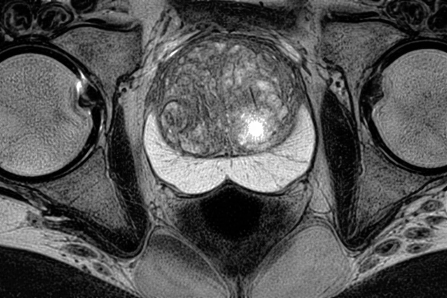
MRI scans provide detailed views of the prostate gland by capturing images of different prostate zones. These images reveal subtle changes in tissue structure that may indicate cancer. Abnormal prostate cells often appear different from healthy tissue when viewed on MRI images.
Radiologists study these images to detect areas where the signal pattern suggests abnormal cell activity. Suspicious areas may appear darker or brighter depending on the imaging technique used during the MRI scan for prostate cancer.
MRI technology can also highlight the shape, size, and boundaries of lesions within the prostate gland. This information helps doctors determine whether the lesion may be cancerous and whether further testing is required.
The PI RADS scoring system is widely used by radiologists to evaluate MRI imaging for prostate cancer. PI RADS stands for Prostate Imaging Reporting and Data System. It provides a standardized method to describe how suspicious a lesion appears on an MRI scan.
This scoring system helps doctors interpret MRI results more consistently and decide whether a biopsy should be performed.
The PI RADS scoring scale includes the following levels.
Higher PI RADS scores usually lead doctors to recommend further evaluation through biopsy or additional imaging.
Prostate cancer diagnosis often involves multiple tests that work together to provide accurate results. PSA testing, MRI scanning, and biopsy each play a different role in evaluating prostate health.
PSA testing measures the level of prostate specific antigen in the blood. Elevated PSA levels may suggest prostate disease, but they do not always confirm cancer. Conditions such as prostate enlargement or infection can also raise PSA levels.
An MRI scan for prostate cancer provides visual information about the prostate gland. It identifies suspicious areas and helps doctors determine whether a biopsy is necessary.
A prostate biopsy involves removing small tissue samples from the prostate gland for laboratory analysis. This test confirms whether cancer cells are present.
PSA Test
MRI Scan
Prostate Biopsy
Together, these tests create a comprehensive approach to prostate cancer screening and diagnosis.
Doctors recommend MRI for prostate cancer screening in several clinical situations. This imaging method provides detailed information about the prostate gland and helps guide further medical decisions.
In many cases, MRI is used when PSA test results show elevated levels. MRI can help determine whether the increase is related to cancer or another prostate condition. It is also helpful when previous biopsy results were inconclusive.
Doctors may recommend prostate MRI screening for the following situations.
MRI imaging for prostate cancer has become an important step before biopsy in many diagnostic protocols.
A prostate MRI scan is a non invasive imaging procedure performed inside an MRI scanner. The process usually takes between thirty and forty five minutes.
Before the scan begins, the patient lies on a movable table that slides into the MRI scanner. The scanner uses magnetic fields to create detailed images of the prostate gland and surrounding tissues.
During the scan, the patient must remain still so that clear images can be produced. In some cases, contrast dye may be injected into a vein to improve visibility of prostate structures.
Steps involved during a prostate MRI scan include the following.
1 The patient lies comfortably on the MRI scanning table
2 The table moves into the scanner tunnel where imaging occurs
3 Magnetic fields capture multiple detailed images of the prostate
4 Contrast material may be used to improve image clarity
5 A radiologist reviews the images and prepares a diagnostic report
This imaging procedure is painless and does not involve radiation exposure.
MRI has significantly improved the accuracy of prostate cancer detection. Advanced imaging methods allow doctors to identify suspicious lesions that may not be visible through other diagnostic tests.
Multiparametric MRI combines different imaging techniques to analyze prostate tissue in detail. These techniques evaluate tissue density, cellular activity, and blood flow patterns inside the prostate gland.
Because of this detailed imaging capability, MRI scans can detect clinically significant prostate cancer more effectively than traditional screening alone. MRI also helps doctors avoid unnecessary biopsies when the imaging results show no suspicious lesions.
Research studies have shown that MRI imaging for prostate cancer improves detection of aggressive tumors while reducing detection of low risk cancers that may not require immediate treatment. This approach helps doctors plan personalized treatment strategies for each patient.
Prostate cancer screening recommendations often depend on age, medical history, and individual risk factors. MRI screening may be recommended for men who show signs of increased prostate cancer risk.
Men who may benefit from MRI screening include the following.
MRI for prostate cancer screening helps doctors evaluate the prostate gland in detail and determine whether further diagnostic testing is necessary.
To understand all available diagnostic options, explore our guide on prostate cancer tests, including PSA, blood, urine, and biopsy.
Can MRI detect prostate cancer without a biopsy?
MRI scans can identify suspicious prostate lesions and estimate the likelihood of cancer. However, a biopsy remains necessary to confirm the presence of cancer cells and determine the exact diagnosis.
Is MRI the best scan for prostate cancer detection
Multiparametric MRI is considered one of the most advanced imaging tests for prostate cancer. It provides detailed prostate images and helps doctors detect aggressive tumors more accurately.
How long does a prostate MRI scan take
A typical MRI scan for prostate cancer takes about thirty to forty-five minutes. The exact duration may vary depending on the imaging protocol and whether contrast material is used.
Is MRI safe for prostate cancer screening
MRI scanning does not use radiation and is generally considered safe for prostate cancer screening. Most patients tolerate the procedure well and experience minimal discomfort.
Can MRI replace prostate biopsy
MRI cannot replace biopsy entirely. It helps identify suspicious lesions and guide targeted biopsy procedures, but tissue sampling is still required to confirm prostate cancer diagnosis.
Do all men with high PSA need MRI
Not all men with elevated PSA require MRI. Doctors evaluate other clinical factors such as age, medical history, and examination results before recommending MRI imaging for prostate cancer.
MRI has become a valuable tool in modern prostate cancer diagnosis and screening. Detailed MRI imaging for prostate cancer allows doctors to detect suspicious lesions, guide biopsies, and evaluate the extent of disease more accurately. When used together with PSA testing and clinical examination, MRI improves the ability to identify clinically significant prostate cancer while reducing unnecessary procedures. Early detection supported by advanced imaging technology plays a key role in improving treatment planning and long-term patient outcomes.
MVR offers trusted and advanced MRI scan services in Dubai for the accurate detection of various health conditions, including prostate cancer. The center uses high-quality imaging technology and skilled medical professionals to deliver precise diagnostic results. Patients benefit from comfortable scanning facilities, efficient reporting, and expert radiology interpretation. MVR also provides affordable screening and diagnostic packages, making advanced MRI imaging accessible for individuals who need reliable and professional diagnostic care in Dubai.
WhatsApp us