
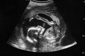
You are pregnant and your doctor recommends an echo for pregnancy. Your first thought is simple. Is this safe for my baby? A prenatal echocardiogram is a detailed scan that checks your baby’s heart between 20 to 24 weeks. It uses sound waves, not radiation, and is guided by medical specialists. If you feel anxious or confused, read on to know what fetal echocardiography involves, why it is done, and what to expect.
Fetal echocardiography is a specialized ultrasound test that closely examines the structure and function of your baby’s heart while still inside the womb. Unlike a routine pregnancy scan that checks overall growth and anatomy, this test focuses only on the heart chambers, valves, blood vessels, and blood flow patterns. It gives a clearer and more detailed view of how the heart is developing.
This prenatal echocardiogram is usually recommended between 20 to 24 weeks of pregnancy. At this stage, the baby’s heart is developed enough for doctors to evaluate its structure clearly. The scan uses high frequency sound waves that create images on a screen. There is no radiation involved, which makes it different from X rays or CT scans. Because of this, fetal echocardiography is considered safe and non invasive.
To learn more about different prenatal tests, read our guide on Pregnancy Scans Explained: Types, Timing, and What to Expect.
Yes, fetal echocardiography is considered safe during pregnancy when performed by trained medical professionals using certified ultrasound equipment. The technology used in this scan is the same as regular pregnancy ultrasound, but it is more detailed and focused on the heart. It does not expose the mother or baby to harmful radiation.
Here are important safety points to know:
A prenatal echocardiogram is performed by experienced specialists who adjust the settings to ensure the safest possible exposure. The scan is done externally on the abdomen, so nothing enters the body. When done correctly, it does not increase the risk of miscarriage or premature labor. Doctors rely on this test because the benefits of early detection of heart conditions are significant for both mother and baby.
Doctors may suggest an echo for pregnancy when there is a higher chance of heart-related issues. While many pregnancies do not require this test, certain risk factors make it important for detailed evaluation.
A fetal echocardiography may be recommended in the following situations:
In these cases, a prenatal echocardiogram provides clarity and helps doctors plan the next steps. Even if there is no clear risk factor, some parents choose to do the test for reassurance. The goal is early identification of any structural problem so that proper care can be arranged before delivery.
The fetal echo scan is done in a similar way to a regular ultrasound, but it takes more time and focuses only on the baby’s heart. The mother lies comfortably on an examination bed while a clear gel is applied to the abdomen. This gel helps the ultrasound probe move smoothly and improves image clarity.
The procedure usually follows these steps:
The test is painless and does not require anesthesia. Sometimes the baby’s position may make it slightly longer, but there is no discomfort beyond mild pressure from the probe. The focus is on capturing detailed heart images for accurate evaluation.
A fetal echocardiography usually takes between 30 to 45 minutes. The duration depends on several factors such as the baby’s position, heart rate, and how clearly the structures can be seen. If the baby is moving frequently, the scan may take slightly longer because the specialist needs clear views from different angles.
In some cases, the doctor may ask the mother to change position or walk briefly to help the baby move into a better position. This is normal and part of ensuring accurate results. Even if the scan takes longer than expected, it remains safe and comfortable. The extra time allows for a thorough assessment of heart development and blood flow patterns.
Most women do not need special preparation before a prenatal echocardiogram. Unlike early pregnancy scans, a full bladder is usually not required at 20 to 24 weeks because the uterus is large enough for clear imaging.
Preparation guidelines include:
You can eat normally before the scan unless your doctor advises otherwise. There is no need for fasting. Knowing what to expect can reduce anxiety and make the experience smoother.
Fetal echocardiography is designed to detect many structural and functional heart problems before birth. Early detection helps parents and doctors prepare for necessary medical care after delivery.
The scan can identify:
While it can detect most major heart issues, it may not identify every minor abnormality. The level of detail depends on fetal position and equipment quality. However, it remains one of the most advanced tools for assessing fetal heart health. Early diagnosis allows coordinated care involving obstetricians and pediatric cardiology teams.
The fetal echocardiography cost in the UAE is generally affordable compared to many other advanced diagnostic tests. In most diagnostic centers, the average price is around 600 AED when performed between 20 to 24 weeks of pregnancy.
The cost may vary slightly depending on the facility, the experience of the specialist, and the equipment used. Some clinics offer pregnancy scan packages that include anomaly scans and echo for pregnancy evaluations. It is advisable to choose a trusted diagnostic center that provides clear pricing and certified medical supervision.
Many parents worry about possible side effects, but fetal echocardiography does not harm the baby when performed correctly. It is based on ultrasound technology, which has been used safely in pregnancy care for decades. There is no exposure to radiation, and the procedure does not interfere with fetal development.
Doctors monitor the intensity and duration of ultrasound exposure carefully. The equipment is designed to follow strict safety guidelines. Because of this, a prenatal echocardiogram is considered safe even when repeated if medically necessary. The reassurance and early detection benefits often outweigh any minimal concerns.
A fetal echo scan offers valuable insight into your baby’s heart health during pregnancy. It is safe, non invasive, and performed between 20 to 24 weeks using sound waves. The information gained from fetal echocardiography helps doctors detect heart defects early and plan appropriate care. Knowing the fetal echocardiography cost in advance, typically around 600 AED in the UAE, also allows families to prepare financially. If your doctor recommends a prenatal echocardiogram, it is usually for added safety and reassurance.
For reliable echo for pregnancy services in Dubai, MVR provides trusted and advanced diagnostic care using high quality ultrasound technology and experienced medical professionals. Every prenatal echocardiogram is performed with focus on accuracy, patient comfort, and detailed reporting. MVR offers affordable fetal echocardiography packages, ensuring accessible screening for expectant parents. Choose a center that values precision, safety, and compassionate care for every stage of pregnancy.
Is echo for pregnancy safe in the second trimester?
Yes. It is usually done between 20 to 24 weeks and uses sound waves, not radiation. When performed by trained specialists, it is considered safe for both mother and baby.
What is the difference between anomaly scan and prenatal echocardiogram?
An anomaly scan checks overall fetal anatomy, while a prenatal echocardiogram focuses specifically on detailed heart structure, blood flow, and rhythm patterns.
How accurate is fetal echocardiography?
Fetal echocardiography is highly accurate for detecting major congenital heart defects when performed by experienced specialists using advanced ultrasound equipment.
Does fetal echocardiography cost a lot in UAE?
The average fetal echocardiography cost in the UAE is around 600 AED, though prices may vary slightly depending on the clinic and specialist expertise.
Can I eat before a fetal echo scan?
Yes. Most patients can eat normally before the scan. Fasting is usually not required unless specifically advised by the doctor.
What happens if a heart issue is found?
If a heart abnormality is detected, doctors may refer you to a pediatric cardiology team to plan monitoring, delivery strategy, and post birth treatment if needed.
WhatsApp us