

Sports injuries and sudden physical strain can sometimes lead to ongoing pain even when X rays show no fracture or bone damage. Ligaments, tendons, muscles, and cartilage are soft tissues that may become stretched, inflamed, partially torn, or severely injured during physical activity or accidents. Read on to learn how soft tissue injury MRI supports detailed evaluation of ligament and tendon damage and why advanced imaging may be recommended when pain or movement problems continue after an injury.
Why Soft Tissue Injuries May Not Appear on X Rays
X rays are mainly used to identify fractures and major bone related injuries, but they do not provide detailed images of ligaments, tendons, muscles, or cartilage. Because of this, a person may continue experiencing swelling, pain, weakness, or limited movement even when X ray results appear normal after a sports injury or sudden strain.
Soft tissue injuries can develop from twisting movements, falls, repetitive physical stress, sudden impact, or overuse during sports and exercise. In some cases, symptoms improve with rest and rehabilitation, while more significant injuries may continue affecting mobility and daily activity. Doctors often consider soft tissue injury MRI when symptoms remain persistent or when physical examination findings suggest deeper tissue damage.
How Ligament and Tendon Injuries Affect Movement
Ligaments and tendons play important roles in joint stability and body movement. Ligaments connect bones to each other, while tendons connect muscles to bones. Injury to either structure may affect flexibility, strength, balance, and joint function.
Soft Tissue Injury MRI for Ligament Damage
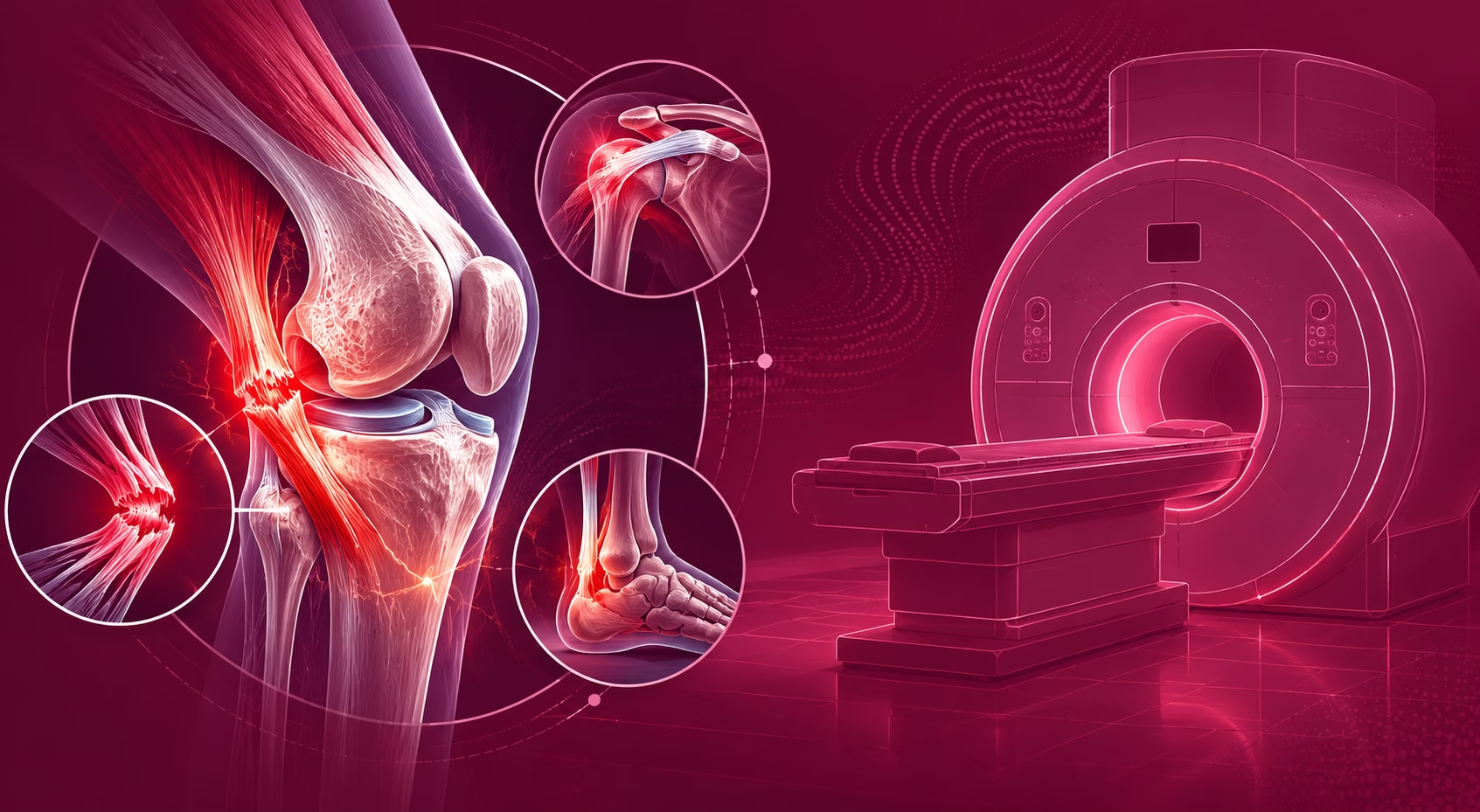
Ligaments may stretch or tear during sudden twisting movements or direct impact injuries. Knee, ankle, shoulder, and wrist ligaments are commonly affected during sports activities. People with ligament injuries may notice swelling, instability, reduced range of motion, or difficulty placing weight on the affected area. Soft tissue injury MRI helps evaluate the severity and exact location of ligament injuries more clearly.
Tendon Damage and Ongoing Pain
Tendons may become inflamed, strained, or torn due to overuse, sudden force, or repetitive stress. Pain often becomes more noticeable during movement or physical activity. Some tendon injuries develop gradually over time, especially in athletes or individuals performing repeated physical tasks. MRI imaging supports detailed assessment of tendon thickness, inflammation, and possible tears.
Muscle and Cartilage Related Symptoms
Soft tissue injuries may also involve surrounding muscles or cartilage structures. Muscle tears can cause weakness and stiffness, while cartilage damage may contribute to joint locking, clicking, or persistent discomfort. MRI helps doctors examine these tissues together for a more complete injury assessment.
How Soft Tissue Injury MRI Supports Detailed Evaluation
MRI, or Magnetic Resonance Imaging, uses magnetic fields and radiofrequency signals to create highly detailed images of soft tissues inside the body. Unlike X rays, soft tissue injury MRI provides clear visualization of ligaments, tendons, muscles, cartilage, and surrounding structures without using radiation.
Situations Where Soft Tissue Injury MRI May Be Recommended
- Persistent pain after sports injuries
- Swelling that continues despite rest
- Reduced joint movement or stiffness
- Suspected ligament tears
- Tendon pain during movement or exercise
- Weakness or instability in joints
- Ongoing symptoms despite normal X ray findings
What Soft Tissue Injury MRI May Help Detect
- Partial or complete ligament tears
- Tendon inflammation or rupture
- Muscle strain or muscle tears
- Joint fluid accumulation
- Cartilage damage
- Soft tissue swelling and inflammation
In many healthcare facilities across the UAE, soft tissue injury MRI is commonly recommended for sports injuries because it provides detailed soft tissue evaluation and helps guide treatment planning more accurately.
Common Areas Evaluated With Soft Tissue Injury MRI
Different joints and body regions may require MRI imaging depending on where symptoms are located and how the injury occurred. Doctors combine symptom history, physical examination, and imaging findings when planning further management.
Knee and Ankle Injuries
Knee MRI is frequently used to assess ligament injuries involving structures such as the ACL, meniscus, or collateral ligaments. Ankle MRI may help evaluate tendon strain, ligament damage, and joint instability after sports related twisting injuries.
Shoulder and Wrist Injuries
Shoulder MRI is commonly used to examine rotator cuff tendons, labral injuries, and surrounding soft tissue inflammation. Wrist MRI may support assessment of ligament damage, tendon irritation, or repetitive strain injuries affecting hand and wrist movement.
Muscle and Sports Related Overuse Injuries
Athletes and physically active individuals may develop muscle tears or overuse injuries from repetitive activity. Soft tissue injury MRI helps evaluate the severity of tissue damage and may assist healthcare providers in planning rehabilitation and return to activity recommendations.
Safety and Preparation During Soft Tissue Injury MRI
Soft tissue injury MRI is considered a non invasive imaging method and does not involve radiation exposure. The procedure is commonly used for both acute injuries and long term sports related pain evaluation.
What Patients Usually Experience During MRI
- Lying still on the MRI scanning table
- Hearing rhythmic machine sounds during imaging
- Wearing ear protection for comfort
- Following breathing instructions during certain scans
- Remaining still to improve image quality
Important Safety Information Before MRI
- Informing staff about implants or medical devices
- Removing metal objects before the scan
- Discussing pregnancy status if applicable
- Mentioning claustrophobia concerns beforehand
- Sharing previous injury reports or imaging results
Choosing the Right Imaging Approach for Sports Injuries
Soft tissue injuries involving ligaments, tendons, muscles, and cartilage may continue causing pain even when fractures are not visible on X rays. Soft tissue injury MRI supports detailed evaluation by providing clear images of soft tissue structures and helping identify injuries that may affect movement and joint stability. Early imaging assessment can support accurate diagnosis, appropriate rehabilitation planning, and safer recovery decisions for individuals experiencing ongoing pain or mobility problems after sports related injuries.