
A 2D ultrasound is one of the most common and safest medical imaging methods used today. It helps doctors see inside your body using sound waves, not radiation. This quick, painless test is often done during pregnancy or for examining internal organs. In just a few minutes, it gives doctors valuable information to help diagnose, monitor, or confirm medical conditions confidently.
What Is a 2D Ultrasound?
A 2D ultrasound, also called a sonography or ultrasound scan, is a diagnostic imaging technique that creates two-dimensional images of the body. It works by sending sound waves through soft tissues and capturing the echoes that bounce back. These echoes are converted into real-time images on a monitor, allowing doctors to see organs, tissues, and blood flow.
Unlike X-rays, a 2D ultrasound does not use radiation. It is considered safe for both adults and unborn babies. Because it is noninvasive and provides quick results, it is widely used for both medical diagnosis and preventive screening.
How Does It Work ?
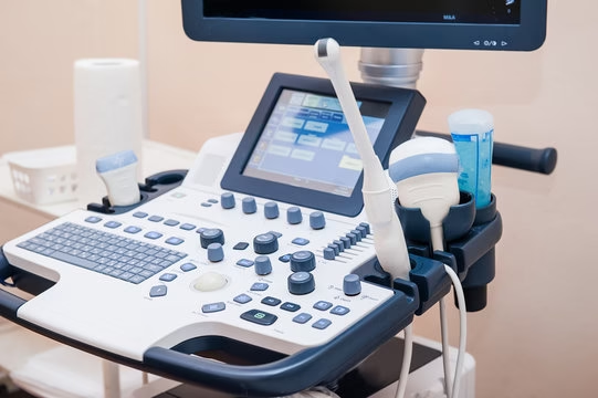
The working principle of a 2D ultrasound is simple yet effective. A small device called a transducer emits high-frequency sound waves into the body. These waves travel through tissues and bounce back when they hit internal structures like organs or bones.
Here’s how the process works:
- The transducer sends out sound waves.
- These waves reflect off internal tissues.
- The returning echoes are captured by the same transducer.
- A computer processes the echoes and converts them into moving images.
These images appear instantly on a monitor, allowing doctors to study the shape, size, and movement of internal organs or a developing fetus.
2D Ultrasound Uses
2D ultrasound is used across many medical areas because of its accuracy, speed, and safety. Some of the most common uses include:
- Pregnancy ultrasound: To confirm pregnancy, estimate gestational age, monitor fetal growth, and check the baby’s position or heartbeat.
- Abdominal scans: To assess organs such as the liver, kidneys, gallbladder, and pancreas.
- Cardiac ultrasound (Echocardiogram): To visualize the heart’s chambers and valves.
- Musculoskeletal scans: To evaluate joints, muscles, and tendons for injuries or inflammation.
- Pelvic ultrasound: To study the uterus, ovaries, and bladder.
In hospitals, diagnostic centers, and clinics, 2D ultrasound is often the first step before recommending more advanced imaging tests.
Why Doctors Order 2D Ultrasound
Doctors recommend a 2D ultrasound for several reasons. It is an essential diagnostic tool that helps detect and monitor conditions without any side effects.
Some common purposes include:
- To monitor fetal anatomy and development during pregnancy.
- To check for cysts, tumors, or internal bleeding.
- To evaluate pain or swelling in soft tissues or organs.
- To guide medical procedures like biopsies or fluid drainage.
- To screen organ function and structure in preventive checkups.
Because it provides instant visual information, 2D ultrasound helps doctors make quick, accurate clinical decisions.
Step-by-Step: What Happens During the Procedure
A 2D ultrasound is a simple, painless process that usually takes 15 to 30 minutes. Here’s what typically happens:
- You will be asked to lie down on an examination table.
- A clear, water-based gel is applied to the skin over the area being examined.
- The sonographer moves a handheld probe (transducer) over the area.
- The probe sends and receives sound waves that create images on the screen.
- The images are captured and stored for a doctor’s review.
There is no recovery time required after the test, and you can return to your normal activities immediately.
Patient Preparation Guidelines
Preparation depends on the type of ultrasound being done. Some scans require fasting, while others may need a full bladder for better imaging.
Common preparation tips:
- Drink plenty of water before a pelvic or pregnancy scan.
- Avoid eating for 6 to 8 hours before an abdominal scan.
- Wear comfortable, loose clothing.
- Remove jewelry or metal accessories.
- Follow your doctor’s instructions carefully before the appointment.
Proper preparation ensures clearer images and more accurate results.
What You’ll Experience During a 2D Ultrasound
During a 2D ultrasound, most patients feel completely comfortable. The procedure is noninvasive and does not cause pain. The gel applied may feel cool on the skin, and you might experience mild pressure as the transducer is moved across your body.
Patients describe the experience as:
- Quick and easy
- Comfortable and stress-free
- Free from side effects or discomfort
After the scan, the gel is wiped off easily, and there is no downtime required.
Benefits & Advantages of 2D Ultrasound
2D ultrasound remains one of the most trusted medical imaging tools because of its safety, speed, and reliability.
Main benefits include:
- Completely safe and uses no ionizing radiation.
- Noninvasive and painless.
- Cost-effective compared to other imaging methods.
- Provides real-time imaging for accurate diagnosis.
- Widely available in hospitals and diagnostic centers.
- Suitable for all age groups, including pregnant women.
Its accessibility makes it a preferred choice for both routine checkups and emergency evaluations.
Limitations & When 2D Isn’t Enough
While 2D ultrasound is effective for many conditions, it has a few limitations. It produces flat images that may not capture all angles or fine details.
Limitations include:
- Reduced clarity in obese patients or those with excessive gas.
- Limited depth for viewing deep structures.
- Difficulty in capturing blood flow details.
- Dependence on operator skill for image quality.
In such cases, doctors may suggest other imaging methods such as 3D ultrasound, Doppler ultrasound, MRI, or CT scan for deeper evaluation.
Risks, Safety & Compliance
2D ultrasound is among the safest diagnostic tools in modern medicine. It uses sound waves instead of radiation, so there is no risk of cell damage.
Medical authorities worldwide recognize its safety record. When performed by trained professionals, it poses no known harmful effects to patients or unborn babies.
Safety guidelines followed include:
- Limiting exposure time to necessary duration.
- Using approved ultrasound frequencies.
- Following recommended scanning protocols.
Regulatory bodies ensure that ultrasound machines meet safety standards, ensuring complete patient protection.
Contraindications or Cautions
Though ultrasound is safe, it might not always provide clear results in certain conditions.
Common limitations or cautions:
- People with obesity may have unclear images due to tissue depth.
- Excessive bowel gas can interfere with sound wave transmission.
- Dense bones may block the ultrasound beam.
In such cases, a doctor may recommend alternative imaging options for better clarity.
Interpreting the Results
After the scan, a radiologist or sonographer reviews the captured images. They analyze structures, shapes, and movements seen in the ultrasound and prepare a report for the referring doctor.
Results are usually available within a few hours or the next day. The doctor then explains what the images indicate and whether further tests are required.
This quick reporting system allows early detection and timely treatment of many health conditions.
Common Findings & What They Mean
The meaning of ultrasound results depends on the purpose of the scan.
Some examples include:
- Pregnancy scans: Show fetal position, heartbeat, and growth progress.
- Abdominal scans: Detect gallstones, liver enlargement, or kidney issues.
- Cardiac scans: Reveal heart chamber size and blood flow patterns.
- Pelvic scans: Identify cysts, fibroids, or reproductive organ abnormalities.
Normal results show well-defined organ shapes and regular movement. Abnormal results might require further imaging or consultation.
Cost, Access & Locations
The cost of a 2D ultrasound depends on the body area being scanned and the location of the diagnostic center. On average, it is one of the most affordable imaging tests available.
You can easily find ultrasound near me searches to locate nearby diagnostic centers or hospitals offering this service. Many clinics offer same-day appointments and results.
Factors affecting cost include:
- Type of ultrasound (abdominal, pelvic, or pregnancy).
- Hospital or clinic location.
- Additional scans or follow-up appointments.
Overall, 2D ultrasound remains a budget-friendly and accessible choice for accurate imaging.
2D Ultrasound Cost in Dubai
The cost of a 2D ultrasound in Dubai varies depending on the type of scan, medical facility, and additional services provided. On average, the price ranges between AED 150 to AED 500 for a single session. Maternity or specialized scans, such as pelvic or transvaginal ultrasounds, may cost slightly higher.
Approximate price range by type:
- General abdominal or pelvic ultrasound: AED 150 – AED 300
- Pregnancy ultrasound (single fetus): AED 250 – AED 400
- Transvaginal ultrasound: AED 300 – AED 500
- Echocardiogram (2D heart scan): AED 350 – AED 600
Factors influencing the cost include:
- Clinic or hospital reputation – premium healthcare centers may charge more.
- Doctor’s expertise – specialist consultations may add to the price.
- Location – central Dubai areas usually have higher rates.
- Additional services – color Doppler, reports, or follow-up consultations.
Most diagnostic centers in Dubai provide packages or online appointment options for convenience.
MVR Diagnostic Clinic in Dubai offers affordable ultrasound services, including 2D ultrasound.
Tips for Patients & Best Practices
To make your ultrasound experience smooth and effective, keep these points in mind:
Helpful tips:
- Follow preparation instructions carefully.
- Arrive early to complete registration and relax before the test.
- Ask questions if you have doubts about the procedure.
- Keep past medical records handy for comparison.
- Inform the sonographer about any discomfort during the scan.
Choosing a reputable sonography clinic with experienced professionals ensures the best quality images and reliable reports.
Choosing 2D Ultrasound for Safe and Accurate Diagnosis
A 2D ultrasound is one of the most trusted diagnostic tools in modern healthcare , safe, quick, and reliable. Whether it’s for pregnancy monitoring, abdominal screening, or heart evaluation, this imaging method provides instant insights without any radiation risk. With its affordability and accuracy, 2D ultrasound continues to play a vital role in early detection, preventive care, and ongoing medical monitoring.
Affordable 2D Ultrasound at MVR Diagnostic Clinic, Dubai
MVR Diagnostic Clinic offers affordable and high-quality 2D ultrasound services in Dubai, ensuring accurate imaging and quick results for patients. Using advanced ultrasound technology and experienced radiologists, MVR provides safe and precise scans for pregnancy, abdominal, pelvic, and general health assessments.